Paper - The Development of Head-Process and Prochordal Plate in Man
| Embryology - 30 Apr 2024 |
|---|
| Google Translate - select your language from the list shown below (this will open a new external page) |
|
العربية | català | 中文 | 中國傳統的 | français | Deutsche | עִברִית | हिंदी | bahasa Indonesia | italiano | 日本語 | 한국어 | မြန်မာ | Pilipino | Polskie | português | ਪੰਜਾਬੀ ਦੇ | Română | русский | Español | Swahili | Svensk | ไทย | Türkçe | اردو | ייִדיש | Tiếng Việt These external translations are automated and may not be accurate. (More? About Translations) |
Hill JP. and Florian J. The development of head-process and prochordal plate in man (1931) J Anat. 65(2): 242-6. PMID 17104317
| Historic Disclaimer - information about historic embryology pages |
|---|
| Pages where the terms "Historic" (textbooks, papers, people, recommendations) appear on this site, and sections within pages where this disclaimer appears, indicate that the content and scientific understanding are specific to the time of publication. This means that while some scientific descriptions are still accurate, the terminology and interpretation of the developmental mechanisms reflect the understanding at the time of original publication and those of the preceding periods, these terms, interpretations and recommendations may not reflect our current scientific understanding. (More? Embryology History | Historic Embryology Papers) |
The Development of Head-Process and Prochordal Plate in Man
- Communicated to the International Congress of Anatomy, Amsterdam, 1930.
By J. P. Hill, D.Sc., F.R.S., and J. Florian, M .D.
From the Department of Anatomy and Embryology, University College, London
(Preliminary Note)
This preliminary note is the outcome of our study of a young human embryo (Dobbin embryo, 960, µ long), which was presented to one of us (Hill) by Dr Roy Dobbin of Cairo. The embryo belongs to the stage characterised by the presence of a head-process with a well-developed chorda-canal.
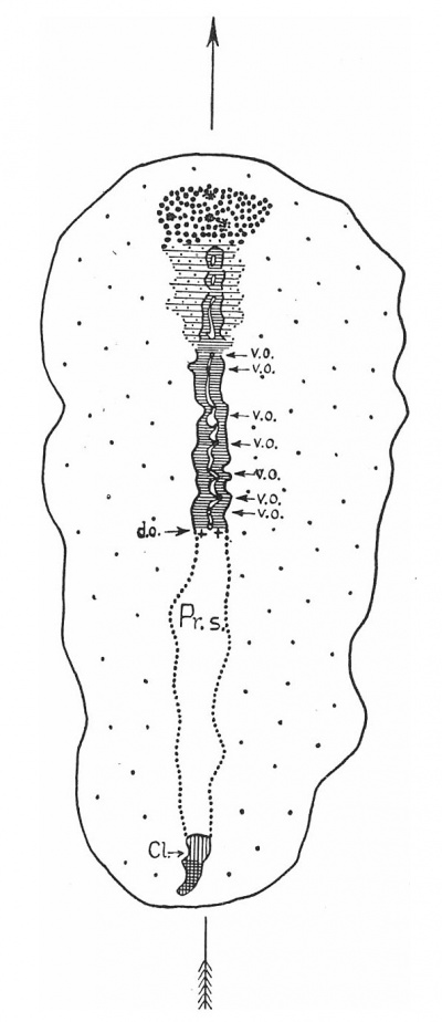
Dr Roy Dobbin of Cairo. The embryo belongs to the stage characterised by the presence of a head-process with a well-developed chorda-canal. In the reconstruction of the dorsal view of the embryonal shield (fig. 1), the axial formation which extends cranially in front of Hensen’s knot is distinguishable into three portions, a caudal portion forming about the caudal half of the entire formation, a very short but broad cranial portion, and between these two, an intermediate portion which narrows back from the latter to pass into continuity with the caudal portion. The caudal portion is formed by a typical chordacanal, with seven ventral openings into the yolksac cavity and a dorsal opening into the amniotic cavity; the cranial portion represents, in our opinion, the region of undoubted prochordal plate, but we find the intermediate region less easy of interpretation. In front of the most cranial ventral opening of the chorda-canal, the head-process continues directly forwards into the intermediate region as a well-marked cylindrical cord. Solid in two sections, it then develops a lumen which extends through six sections before it ends blindly. In front of the just-mentioned canal, two additional lumina, also closed but of much shorter extent than the first, make their appearance in the median cell-cord. It differs from the more caudal portion of the head-process in two important respects: (1) it is accompanied on both sides by thickened bands of mesoderm which are in continuity peripherally with the forward extensions of the primitive streak mesoderm and which we shall distinguish as the “lateral mesodermal bands ”; (2) below it, the endoderm is for the most part distinct, and there is clear evidence that in quite a number of sections it is separated from the median cell cord by mesoderm cells. Cranially the median cell cord and the lateral mesodermal bands pass over into a single, very thick and broad mass, composed of mesodermal cells with which the underlying endoderm is indistinguishably fused. We regard this mass as representing mesoderm derived from the prochordal plate thickening of the endoderm. There are present in it numerous chromatophilic granules similar to those originally described by Bonnet in the prochordal plate (“Erganzungsplatte”) of the Dog. '
The precise developmental relations between this undoubted prochordal plate region and what we have distinguished as the intermediate region of the axial formation obviously cannot be determined from the study of one specimen, and can only be finally settled by the study of a close series of well preserved embryos. We have made, however, a very careful study of the intermediate region in our specimen and have examined the corresponding region in various other mammalian embryos, of which an early presomite embryo of Loris (Loris 49 of the collection made by Prof. A. Subba Rau) has proved specially instructive, and the interpretation we have reached is that it represents the cranial segment of the head-process which has undergone differentiation into a median cell cord, the chorda-process, which is in course of becoming luminated and two lateral mesodermal bands, representing socalled “gastral or archenteric” mesoderm. We have no evidence that the prochordal plate participates in the formation of the head-process, and consequently regard that plate~as a purely prochordal structure. It is possible, however, that the head-process secondarily grows into the region of the prochordal plate, and that there is an overlap (perhaps only temporary) between the two where they pass into continuity. But even if this is the case, the prochordal plate need not necessarily be regarded as participating in the formation of the chorda-canal, although it might underlie its most cranial segment. We leave this question of overlap quite open, and only call attention to Rossenbeck’s remark that the endoderm underneath the most cranial part of the chorda-canal in embryo Peh.1-Hochstetter resembles the endoderm of the “Erganzungsplatte.” The view of Ingalls, that the region we have distinguished as “intermediate,” as well as the region we have regarded as the prochordal plate, together constitute the “completion plate” (“Erganzungsplatte”), we cannot accept. Nor can we accept the view of Rabl that the prochordal plate is simply the most cranial part of the head-process which has pushed forwards into the lecitophor. In our opinion, this View is .devoid of any basis of fact, Hill and Tribe (1) having demonstrated the existence in the dog of a mesoderm-producing area in the endoderm (i.e. a prochordal plate) long before the appearance of the other axial structures.
Amongst the described human embryos there are two in which the conditions of the head-process and prochordal plate resemble those in our specimen: the Peh.1-Hochstetter embryo (described by Rossenbeck (3)) and the embryo No. 1 of the Department of Anatomy of Western Reserve University, described by Ingalls (2). According to the measurements of the authors, the former ought to be 1-4 mm. long, the latter 1-8 mm.; but it is evident that the difference in their length is due partly to the adoption by the authors of different points of measurement. We have measured the distance between the cranial margin of the prochordal plate and the cranial margin of the cloacal membrane and find that this distance is in Ingalls’ embryo about l~40 mm., in Rossenbeck’s embryo 1-36 mm., so that there is practically no difference in length between these two specimens. The primitive streak in Ingalls’ embryo is stated to be 0-65 mm. long, in Rossenbeck’s specimen about 0-66 mm. The most important difference between them seems to be that in the Ingalls’ embryo there are three ventral openings in the proper chorda-canal, whilst Rossenbeck found only two sections in two different regions of the chordacanal of his embryo where a ventral opening was apparently forming. This difference seems to be of no significance for the estimation of the developmental stage of the specimens; the Dobbin embryo possesses seven ventral openings in the proper chorda-canal (two of them extending through several sections), nevertheless it is certainly younger than Rossenbeck’s embryo, the difference in the length of the two being nearly 0-5 mm. It is, therefore, clear that there is great individual variation in this respect and that the disappearance of the ventral wall of the chorda-canal cannot be used alone in the determination of the stage of development of a particular specimen.
The differences in the axial structures under consideration in the Dobbin embryo and that of Ingalls are relatively slight. In the Dobbin embryo, as we have stated, there are three closed lumina in the intermediate axial region of the head-process. In the Ingalls embryo, instead of three cavities, there is one continuous canal in this same region (“completion plate” according to Ingalls, head-process according to our interpretation) which possesses a ventral opening of its own, close to its transition into the chorda-canal proper. It may be suggested that the three separate lumina in the Dobbin embryo would in the course of further development join together to form a single canal, similar to that in the Ingalls embryo.
Apart from the difference in the number and size of the ventral openings, it would seem that Rossenbeck had to deal with very similar structures in his embryo so far as can be judged from his description and figures. We can recognise in the axial formation a caudal chorda-canal with a continuous lumen, possessing two very indistinct ventral openings. In front of this is a region corresponding to our intermediate region in which there is present a median chorda-canal and two thickened bands of mesoderm more or less symmetrically disposed on either side of the latter and broader than those in our specimen. According to Rossenbeck, the lumen of the chorda-canal of this region is directly continuous with that of the caudal portion of the canal. It is worthy of note that, whilst we interpret the lateral mesodermal bands as derivatives of the head-process, Rossenbeck regarded them as formed by the mesoderm of the “Erganzungsplatte.” Finally there is present a prochordal plate region, the precise limits of which we are unable to determine from Rossenbeck’s description.
In respect of the differentiation of the head-process in the intermediate region, we may regard our Dobbin embryo as illustrating the stage in which the chorda-process is in process of acquiring a lumen, inasmuch as the latter is still discontinuous and represented by three isolated portions. In the Rossenbeck embryo, it possesses a lumen which is continuous behind with that of the caudal portion of the chorda-canal, whilst in the Ingalls embryo, the lumen already opens into the yolk-sac cavity just in front of the junction of the two regions of the canal.
In this short preliminary note we need only mention three younger embryos, two of them (Bi. 24 and No. 1285 of the collection of the Department of Anatomy of the University of Manchester) as yet undescribed, the third described by Stieve (4) (embryo “Hugo”). The embryo Bi. 24 was presented by Dr 0. Bittmann of Brno to Florian, and is distinctly more differentiated than the embryo “Hugo.” No. 1285 of the Manchester collection was generously handed over to us for description by Prof. J. B. S. Stopford.
The “Hugo” embryo (length 635p. according to our new reconstruction, the median plane of which makes an angle of 64° with the sectional plane) is the earliest human embryo described in which the head-process is clearly developed. It is very short and very indistinctly delimited laterally from the forward prolongations of the primitive streak mesoderm. So far as can be judged from the figures in Stieve’s paper, there is an area in front of the undoubted head-process, where the endoderm is not distinguishable from an axial accumulation of more or less loosely arranged mesoderm cells. With this accumulation, the cranial end of the head-process appears to be continuous, but whether or not this area represents a prochordal plate, we are unable to determine with certainty.
In the very well preserved embryo Bi. 24 (length 620 In), there is a definite but short head-process, the caudal part of which is accompanied by forward prolongations of the primitive streak mesoderm from which it is clearly distinguishable. The cranial part of the head-process is not accompanied by primitive streak mesoderm and passes over in front into an area of thickened endoderm, which we regard as the prochordal plate. It is of comparatively small extent and is not yet very active in mesoderm production.
In the Manchester embryo (length 870 p.) the head-process is still quite short, but it is thicker than that of Bi. 24, whilst the prochordal plate region appears to be more extensive than in that embryo.
Our observations support the following conclusions: The head-process which arises as a forward growth from Hensen’s knot and is accompanied by forward extensions of the primitive streak mesoderm becomes distinguishable during development (embryos “Dobbin,” “Rossenbeck,” “ Ingalls”) into two portions, a caudal part which early assumes the form of a typical chorda-canal and a cranial part which undergoes differentiation in the caudo-cranial direction into a median chorda-process (later transforming into a chorda-canal) and two lateral mesodermal bands (“ gastral or archenteric ” mesoderm). In all the stages We have studied the head-process passes into continuity at its cranial end with the mesoderm of the prochordal plate. Those stages have not furnished any definite evidence of a participation of the prochordal plate in the formation of the chorda-canal.
References
(1) HULM and TRIBE (1924). Quart. J. Micro. Sci. vol. LXVI.
(2) INGALLS(1918). CarnegieContrib.toEmbryology,vol.VU,No.23.
(3) ROSSENBECK(1923). Zeit8chr.f.Anat.u.Entwick.Bd.LXVIII.
(4) STIEVE(1926). Zeitschr.f.mikr.-anat.For8chung,Bd.vu.
Hill JP. and Florian J. The development of head-process and prochordal plate in man (1931) J Anat. 65(2): 242-6. PMID 17104317
Cite this page: Hill, M.A. (2024, April 30) Embryology Paper - The Development of Head-Process and Prochordal Plate in Man. Retrieved from https://embryology.med.unsw.edu.au/embryology/index.php/Paper_-_The_Development_of_Head-Process_and_Prochordal_Plate_in_Man
- © Dr Mark Hill 2024, UNSW Embryology ISBN: 978 0 7334 2609 4 - UNSW CRICOS Provider Code No. 00098G


