Paper - Some observations on the roof of the primordial human cranium (1923)
| Embryology - 26 Apr 2024 |
|---|
| Google Translate - select your language from the list shown below (this will open a new external page) |
|
العربية | català | 中文 | 中國傳統的 | français | Deutsche | עִברִית | हिंदी | bahasa Indonesia | italiano | 日本語 | 한국어 | မြန်မာ | Pilipino | Polskie | português | ਪੰਜਾਬੀ ਦੇ | Română | русский | Español | Swahili | Svensk | ไทย | Türkçe | اردو | ייִדיש | Tiếng Việt These external translations are automated and may not be accurate. (More? About Translations) |
Fawcett E. Some observations on the roof of the primordial human cranium. (1923) J Anat. 57(3): 245-250. PMID 17103974
| Online Editor |
|---|
| This historic 1923 paper by Fawcett describes the development of the human cranium (skull). Also describes a Tatusia embryo, an armadillo.
See also:
|
| Historic Disclaimer - information about historic embryology pages |
|---|
| Pages where the terms "Historic" (textbooks, papers, people, recommendations) appear on this site, and sections within pages where this disclaimer appears, indicate that the content and scientific understanding are specific to the time of publication. This means that while some scientific descriptions are still accurate, the terminology and interpretation of the developmental mechanisms reflect the understanding at the time of original publication and those of the preceding periods, these terms, interpretations and recommendations may not reflect our current scientific understanding. (More? Embryology History | Historic Embryology Papers) |
Some Observations on the Roof of the Primordial Human Cranium
By Professor Fawcett, M.D.
Introduction
It has been the custom for some time to accept the views expressed by Bolk (Petrus Camper, 2 Deel, pp. 315 etc.) which were based on results obtained from specimens stained in bulk by Van Wijhe’s bulk stain and differentiated.
By this method results were obtained so much at variance with what obtains in vertebrates other than man, and so different from anything obtained in the usual way in man, that in 1910 I ventured to say: “ I must confess the appearances in his figures scarcely explain what is seen in this cranium.” Macklin in 1921 in a description of “The skull of a Human Fetus of 43 millimetres greatest length,” is more precise and states that Bolk has been led into quite a number of errors through the method used. It is only fair to say that Bolk himself does not place implicit faith in the methods, ‘for he says on p. 322, “In der mitte der occipitalregion bleibt unterhalb des Knorpelringes ein Feld iibrig das nicht knorpelig Verschlossen wird, wenigstens das von mir cmgewendete Verfahrein ist nicht in Stande einen vollkommenen lmorpeligen Verschluss ans Licht zu fuhren.” ‘(The italics are mine.)
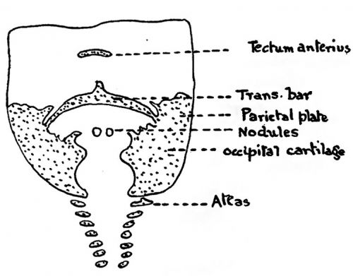
Bolk illustrated his communication by four coloured figures which represent different ages in sequence. In fig. 1 one sees from below upwards, cartilage centres in the cervical vertebrae, separate on the two sides but connected together dorsally by a spinal membrane; above the vertebrae on each side one sees cartilage in a somewhat broad sheet forming-what appears to be the side of a foramen magnum, and connected together about their middle by a narrow bar of cartilage from which a small process ascends. Below this bar is an uncoloured area, Bolk’s spino-occipital membrane, which is continuous with the spinal membrane below and contains two small nodules of cartilage. Above the transverse bar above mentioned is a transversely placed separate mass of cartilage.
Fig. 1. (after Bolk)
Bolk regarded the spino-occipital membrane as reaching up to the transverse bar of cartilage which connects the two side walls of cartilage together, and he rightly said that this transverse bar has nothing to do with bounding the foramen magnum. He did not actually realise that the conditions appearing in his specimen are entirely due to the method used, for all that part lying between the two small nodules and the transverse bar of cartilage is actually cartilage undergoing calcification and therefore has lost its stain during differentiation unless pcrchance it never was stained. Hence the upper segment of the spino-occipital membrane is non-existent.
It is hoped that this communication. will show that there are in reality three tecta in the vault of the cranium, which may be named from behind forwards tectum posterius or tectum occipitale, tectum intermedium or tectum parietale posterius and tectum anterius or tectum parietale anterius.
Fig. 2. A combination of appearances at about 30 mm. C.R. length. Human, viewed from behind.
Text fig. 2 shows the condition of affairs in the chondro-cranium at about the 27 millimetre crown-rump stage. The tectum posterius is seen as a broad band connecting the occipital cartilages above the foramen magnum; from its upper border in the mid-line a short ascending process projects and a broader but short process projects downwards——the processus descendens. These processes were described by me in 1910. Laterally the tectum merges into the corresponding supraoccipital, which is separated incompletely from the parietal plate by an oblique row of unchondrified areas usually associated With the auditory capsule. These areas appear as foramina in the model. They lie along the occipito-parietal groove of Ma.cklin. If they be followed up they lead to a notch situated in the upper edge of the wall of cartilage formed by the supraoccipital cartilage and the parietal plate — Macklin has named this notch the incisura occipito-parietalis. Anterior to the supraoccipital cartilage is the parietal plate (lamina parietalis or lamina supracapsularis). This I think should be described as consisting of an anterior and a posterior part separated by a deep notch. The posterior part is directed upwards and inwards above the outer edge of the supraoccipital cartilage. It either ends freely or is connected with the corresponding spot on the opposite side by a narrow bridge of cartilage - the tectum intcrmedium or parietale posterius. This tectum has only a very short life. It is not present at the 27 mm stage and it has disappeared at the 32 mm. stage. It does not take part in the formation of the interparietal bone and it is not brought out in any of Bolk’s figures.
The anterior part of the parietal plate Varies somewhat in form with age but is well developed at the 30 mm. stage. As it rises in the cranial wall it broadens antero-posteriorly and posteriorly is prolonged as a short angular projection. Stretched between these posterior processes is a chain of three pieces of cartilage, viz. a median and two lateral; these constitute the tectum anterius or tectum parietale anterius.
The two lateral pieces disappear before the 48 mm. stage, but at this stage the median piece still persists. The tecta are thus:
(a) Tectum occipitale or posterius.
(b) Tectum parietale posterius or tectum intcrmedium.
(c) Tectum parietale anterius or tectum anterius. So far as I know only the tectum posterius is converted into bone. Macklin shewed that at the stage described by him, viz. 43 mm., there was an area of calcified cartilage in the occipital tectum and the adjacent supraoccipital eartilages which assumed the form of a butterfly with outstretched wings. In my own specimens at the 27 and 32 mm. stages the same thing occurs. In fig. 2 the calcified area is shown. This area is the area which Macklin has stated is calcified and therefore has either not stained or has lost the stain during differentiation.
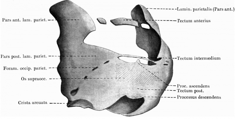
It is easy to see that if the stain were lost from this area two cartilaginous bars would appear to cross the mid-line from side to side and that from the upper bar a processus ascendens would project upwards and from the lower a processus descendens would project downwards into the foramen magnum. The bars would appear to be separated by a clear unstained area, which Bolk thought to be unchondrified. Further as age advances two important changes occur. The processus descendens divides into two processes (fig. 3), and both calcification and ossification so occur in and over the root of these two processes that when viewed from behind (fig. 4), the appearance given in the model at this stage, Viz. 48 mm., is as if there were two separate nodules of cartilage lying in the interval between the two occipital eartilages bounding the iiilcisura posterior of the foramen magnum. The bone which covers the supraoccipital cartilage and tectum occipitale is of a complex nature for it is partly perichondrial and partly dermal (fig. 5). If now figs. 3 and 4 be compared with one another and with fig. 1 which is after Bolk, it can easily be seen how Bolk was led into error. Fig. 3 represents the tectum posterius as viewed from the caval aspect. It will be seen that a bar of cartilage stretches from side to side sending upwards a single process, the processus aseendens, and a process which projects downwards and divides into two. It will be seen that a calcified area, shaped not unlike the butterfly area described by Macklin and marked on the figure by cross-hatching, so modifies the appearance of this deep bar of cartilage as to divide it into an upper bar and two lower nodules connected together above, at their bases, by a narrow band of cartilage. If the same region be now viewed from behind (fig. 4) perichondrial and mixed bone have so extended downwards over the processus deseendens as to hide from view even the roots of the two processes and they appear as isolated nodules. Macklin it was who foreshadowed such an appearance, for he says, p. 66, “It is of interest, however, that this upper margin does remain uncalcified and unossified for such a long period for, as Bolk has shown, it is present throughout his series of four human chondro-crania. In the last, however, it is becoming thinner and more attenuated. I would suggest that this cartilaginous edge may be retained to favour growth of the supraoceipital bone. A similar reason may underlie the persistence of the twin nodules of cartilage which are present in the apex of the superior occipital incisure in Bolk’s preparations, and which he states agree in position in one case at least, with the bones of Kerekring. These nodules are apparently (in Macklin’s model) in the same position as the processus desccndens. It seems to me quite likely that they are not isolated masses of cartilage situated in membrane, but that they are connected with the calcified cartilage of the tectum above.”
Fig. 3. Hinder part of vault of chrondrocranium of human 48 mm C.R. embryo. Here the tectum posterius appears in its full development giving off in the upward direction the processus ascendens and in the downward direction a processus descendens which divides into two. The stippled parts represent uncalcified cartilage, the cross-hatched part is cartilage in process of calcification.
Fig. 4. Reconstruction of the vault of the human chrondrocranium at the 48 mm stage from behind shewing how the processus descendens appears as two isolated cartilages projecting into the incisura posterior and superior of the foramen magnum. Pars canalic. caps. and.
Fig. 5. Model of chondrocranium of 10 mm Tatusia embryo shewing especially the trabeculse diverging behind and giving attachment to the rectus eye muscles; the preoptic limb of the ala orbitalis is not yet laid down. The ala hypochiasmatiea is the posterior free extremity of the trabecula. of its side.
Fig. 5 shews the central stem of the base of the skull in Tatusia at the 10 mm stage. It consists of a chordal segment and a prehypophyseal part. This latter I have usually termed for topographical reasons the pars interorbito-nasalis. In Tatusia this part seems to bifurcate behind and each limb then receives the pre- and post-optic limb of the ala orbitalis from above. The latter being in ontogeny the first to reach the corresponding limb of the pars interorbito-nasalis. That part of each limb of the pars interorbito-nasalis which lies on the floor of the optic foramen is the ala hypochiasmatica and from it the rectus system of eye muscles takes origin. In fig. 5 the muscular origin is shewn, only the post optic limb of the ala orbitalis has joined the pars interorbito-nasalis, but the site of the anterior limb is indicated by a dotted line. There is no lamina hypochiasmatica at this stage. This only appears at the 17mm stage when the two limbs of the pars interorbitonasalis fuse together. The ala hypochiasmatica may thus be looked upon as the edge of the corresponding cornu of the pars interorbito-nasalis which lies below the optic foramen. Should the pars interorbito-nasalis be regarded as the equivalent of the trabeculae, then the alae hypoehiasmaticae would be the fore ends of the posterior cornua of the same.
The figures I have submitted prove that Macklin’s conjecture is correct and that Bolk’s View based on a faulty method is no longer tenable.
My thanks are due to my colleague, Mr D. C. Rayner, for the embryo, and to my assistant, Miss G. R. Llewelyn, for ungrudging help in cutting out wax-plates.
References
BOLK. Petrus Camper, pp. 315-27.
MACKLIN. Contributions to Embryology, No. 48, from Publication 273. Carnegie Institution of Washington, pp. 57-103,
FAWCETT. Journal of Anat. and Physiol. Vol. XLIV, pp. 303-11.
Cite this page: Hill, M.A. (2024, April 26) Embryology Paper - Some observations on the roof of the primordial human cranium (1923). Retrieved from https://embryology.med.unsw.edu.au/embryology/index.php/Paper_-_Some_observations_on_the_roof_of_the_primordial_human_cranium_(1923)
- © Dr Mark Hill 2024, UNSW Embryology ISBN: 978 0 7334 2609 4 - UNSW CRICOS Provider Code No. 00098G


