Paper - Human monochorial twin embryos In separate amnions
| Embryology - 27 Apr 2024 |
|---|
| Google Translate - select your language from the list shown below (this will open a new external page) |
|
العربية | català | 中文 | 中國傳統的 | français | Deutsche | עִברִית | हिंदी | bahasa Indonesia | italiano | 日本語 | 한국어 | မြန်မာ | Pilipino | Polskie | português | ਪੰਜਾਬੀ ਦੇ | Română | русский | Español | Swahili | Svensk | ไทย | Türkçe | اردو | ייִדיש | Tiếng Việt These external translations are automated and may not be accurate. (More? About Translations) |
Potter JC. Human monochorial twin embryos In separate amnions. (1927) Anat. Rec. 34: 253-257.
| Online Editor | |||
|---|---|---|---|
| This historic 1927 paper by Potter describes development of monozygotic twins amnion.
|
| Historic Disclaimer - information about historic embryology pages |
|---|
| Pages where the terms "Historic" (textbooks, papers, people, recommendations) appear on this site, and sections within pages where this disclaimer appears, indicate that the content and scientific understanding are specific to the time of publication. This means that while some scientific descriptions are still accurate, the terminology and interpretation of the developmental mechanisms reflect the understanding at the time of original publication and those of the preceding periods, these terms, interpretations and recommendations may not reflect our current scientific understanding. (More? Embryology History | Historic Embryology Papers) |
Human Monochorial Twin Embryos In Separate Amnions
J. Craig Potter
Anatomical Laboratory, School of Medicine and Dentistry, University of Rochester
2 figures (1927)
Introduction
The rarity of recorded human twin embryos, especially those with a single chorion, has led the author to report this specimen. Although the anatomy and physiology of double monsters and of fetal and adult twins have been given volumes, and complete theoretical explanations of their formation have been proposed, published specimens of twins in embryonic stages to confirm these theories are rare, even in the lower mammals. Corner ( ’22) collected these and added three pig embryos of his own. Human twin embryos of the earlier months are even more rare. Streeter (’19) reported that in the Carnegie collection there were only forty—three specimens, of which all but two were over 5 mm. in length. Arey (’22 a, ’22 b, ’23 a, ’23 b, ’23 c), in a comprehensive series of papers, has gathered and analyzed the cases of monochorionic twins found in tubal pregnancy. Such early embryos are particularly important, since they give information in regard to the method by which twins are presumably derived from a single ovum and the time at which this occurs. As pointed out by Corner, if the embryonic area splits to form twins before the amnion is formed, each twin will lie in a separate amnion surrounded by a single chorion; but if separation occurs after the amnion is formed, the twins will lie in a common amniotic cavity. Of two early specimens reported by Arey, one is of the first type, the other is of the second. The specimen to be described in the present paper is a little older than Arey’s specimens and is of the type in which there are two amniotic cavities, indicating division of the embryonic area at a Very early stage of development, bel7ore formation of the amnions.
Clinical History
Mrs. G. S. C., white, age 23, was admitted to the Rochester General Hospital, with the complaint of vaginal bleeding, while the author was on service, on April 1-}, 1926. Her family history in regard to twins is unknown. Three years previous she had had a miscarriage at three months and two years ago another at two months. Her menstrual periods started at fourteen years, were regular every twenty-eight days, and lasted six: days. During some periods she passed clots. Her last. regular period was 011 February 15, 1S)2(i. "From )1 arch 20, 1.9265, she flowed ten On April 4, 19%, she again started flowing. This continued until the 12th, when clots were passed. When admitted, on April 14th., there was tenderness over the symphysis, the uterus was enlarged to twice normal size, and there was a small mass, slightly larger than an ovary, outside the uterus in the region of the right cornu. On the 15th of April she passed. the prod» nets of conception. This consisted of a single chorion about 8 cm. in diameter which had undergone some maceration. On one surface a portion of the chorion was torn away over an area of about 1 X 2 cm. Though this rent the unruptured amnion bulged slightly. On cutting‘ the amnion, twin embryos were seen lying in apparently separate amniotic sacs. However, before further examination was made, the whole specimen was fixed in '10 per cent formalin for more accurate study.
Because of the retained membranes, a dilatation and curettage was done, which was followed by an uneventful recovery. On the 22nd of April, there was no longer a palpable mass in the region. of the right cornu.
Anatomical Description
When the specimen was thoroughly fixed, it was studied with the following points in mind: the question of separate amnions, separate yolk stalks, the topography of the viscera (because of the well—known fact that twins often have situs inversus viscerorum), and, lastly, the sex of the embryos.
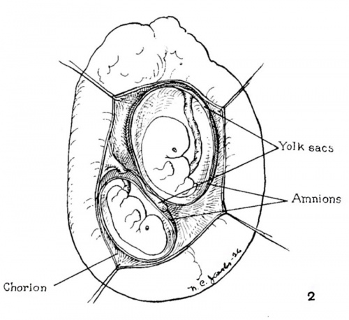
When the chorion was drawn apart in the region of the rent, two distinct amniotic sacs were found. These were gently loosened and found to be absolutely separate through their entirety and arranged so that the attachments of the two yolk stalks lay at different regions of the chorion. An embryo was present in each amniotic sac (fig. 1).
The amnions were then opened and the embryos measured, studied externally, and the yolk stalks observed. The larger embryo measured 19 mm. crown-rump length, the smaller 18.3 mm. The anterior thoracic wall of the smaller embryo was macerated to such a.n extent that the heart protruded. The yolk stalks were separate throughout. That of the smaller embryo could easily be followed down through the cord past its attachment to the chorion and out into the chorionic cavity (fig. 2). The stalk of the larger was traced through the cord to its attachment, but at that point it became blended with, and attached to, the amniotic membrane and did not end in a free bulbous extremity in the chorionic cavity, as did the yolk sac of the smaller embryo.
The study of the viscera was made first in regard to the hearts, after dissecting away the anterior chest wall. Both hearts had been somewhat affected by pressure in fixing, but were definitely located to the left of the midline, with the ventricles pointed to the left.
Serial sections were then made and the viscera. studied under the microscope. The spleens and stomachs were located on the left side in both embryos, but maceration had proceeded so far that it was impossible to determine the sex of the embryos.
The specimen is preserved as no. 60 of the embryological collection of the University of Rochester, School of Medicine and Dentistry.
Discussion
Arey suggested that an altered endometrium might cause intra-uterine twins, in View of the work of Stockard (’21), who showed that in fishes a changed environment would cause monsters and twins. Arey was led to this suggestion because he found that monochorial twins were fifteen times as frequent in tubal pregnancy is bichorial, if the intrauterine ratio was taken as the normal. This, he thought, was possibly due to a disturbed nutritive exchange, the result of previous inflammation, which, as Mall (’15) showed, so frequently precedes ectopic pregnancy. There are three points which make us suspect that the endometrium of the present patient may have been abnormal. She had had three miscarriages, although her uterus was in good position; since her first miscarriage she often passed many clots during her periods; and, finally, there is some suspicion that one or more or these miscarriages were induced, which by leaving infection would injure the endometrium.
The mass found before abortion, which apparently disappeared afterward, was probably a corpus luteum cyst.
Summary
This is a description of early human monochorial twin embryos With separate amnions and yolk stalks, having normal visceral arrangement, in which it was impossible to determine the sex because of maceration.
Bibliography
AREY, L. B. 1922 Chorionic fusion and augmented twinning in the human tube. Anat. Rec., vol. 23, pp. 253-262.
- 1922 Direct proof of the monozygotic origin of human identical twins. Anat. Rec., vol. 23, pp. 245-248.
- 1923 The cause of tubal pregnancy and tubal twinning. Am. Jour. Obstet. and Gynec., vol. 5, pp. 163-167.
- 1923 Tubal twins and tubal pregnancy. Surg., Gynee. and Obst., vol. 36, pp. 803-810.
- 1923 Two embryologically important specimens of tubal twins. Surg., Gynec. and Obst., vol. 36, pp. 407-415.
CORNER, G. W. 1922 The morphological theory of monochorionic twins, as illustrated by a series of supposed early twin embryos of the pig. Johns Hopkins Hosp. Bull., vol. 33, pp. 389-392.
MALL, F. P. 1915 On the fate of the human embryo in tubal pregnancy. Carnegie Institution of Washington, Contribution to Embryol., vol. 1, no. 1, pp. 1-104.
STOCKARD, C. R. 1921 Developmental rate and structural expression. Am. Jour. Ant., vol. 28, pp. 115-278.
STREETER, G. L. 1919 Formation of single—ovum twins. Johns Hopkins Hosp.Bull., vol. 30, pp. 235-238.
Cite this page: Hill, M.A. (2024, April 27) Embryology Paper - Human monochorial twin embryos In separate amnions. Retrieved from https://embryology.med.unsw.edu.au/embryology/index.php/Paper_-_Human_monochorial_twin_embryos_In_separate_amnions
- © Dr Mark Hill 2024, UNSW Embryology ISBN: 978 0 7334 2609 4 - UNSW CRICOS Provider Code No. 00098G


