Paper - Development of the spleen
| Embryology - 28 Apr 2024 |
|---|
| Google Translate - select your language from the list shown below (this will open a new external page) |
|
العربية | català | 中文 | 中國傳統的 | français | Deutsche | עִברִית | हिंदी | bahasa Indonesia | italiano | 日本語 | 한국어 | မြန်မာ | Pilipino | Polskie | português | ਪੰਜਾਬੀ ਦੇ | Română | русский | Español | Swahili | Svensk | ไทย | Türkçe | اردو | ייִדיש | Tiếng Việt These external translations are automated and may not be accurate. (More? About Translations) |
Radford M. Development of the spleen. (1908) J Anat Physiol. 42: 288-301.
| Online Editor |
|---|
| This 1914 historic paper describes spleen development in the embryo. See links below for the modern notes.
|
| Historic Disclaimer - information about historic embryology pages |
|---|
| Pages where the terms "Historic" (textbooks, papers, people, recommendations) appear on this site, and sections within pages where this disclaimer appears, indicate that the content and scientific understanding are specific to the time of publication. This means that while some scientific descriptions are still accurate, the terminology and interpretation of the developmental mechanisms reflect the understanding at the time of original publication and those of the preceding periods, these terms, interpretations and recommendations may not reflect our current scientific understanding. (More? Embryology History | Historic Embryology Papers) |
Development Of The Spleen
By Marion Radford,
Birmingham University.
I. Introductory
The question of the origin and early development of the spleen has been the object of researches since the year 1867. A considerable amount of literature has accumulated since that time, in which the authors dealing with different classes of Vertebrata, and even those who have studied the question in the same animals, have arrived at widely differing conclusions.
Some have believed the spleen to be derived from the mesenchyme of the dorsal mesentery, with or without the participation of the coelomic epithelium; some consider the coelomic epithelium to take the most important part in its development. Some, again, statethat it arises together with the pancreas, while others believe it to have its origin in the endodermal epithelium of the alimentary canal.
1. The origin from the pancreas has been supported by Gotte in 1867 and Schenk in 1873, both of whom were dealing with birds. Kupffer in 1892 claimed to have found the spleen in ganoids arising as a diverticulum of the pancreas.
Woit in 1897 states that in birds and in Siredon it arises “in genetic relation with the pancreas,” but does not consider this to be the case with Rana. Glas (1900) describes a “lieno-pancreas” in Tropidonotus, with a duct leading into the intestine. Orru, Efisio (1902), also gives the pancreas a share in the formation of the spleen. I may quote in this connection from Schenk (iv. p. 627). His personal observations apply only to the chick and mammals.
“Das Mesenchym, welches die Hauptmasse der fiir das Pankreas bestimmten Mesodermgebilde gibt, hangt innig mit jenem Theile des Mesenchyms zusammen, welches die Anlage der Milz bi1det.” He considers that the spleen arises together with the pancreas, but is part of the mesoderm into which the pancreatic diverticulum grows. Kupffer, on the other hand (quoted by Laguesse), definitely gives the spleen an endodermal origin. In the Sturgeon he found four pancreatic buds, two dorsal and two ventral. The dorsal posterior diverticulum, according to him, divides into three branches: of these the right continues pancreatic in structure, the middle one forms the sub-chordal lymphoid tissue, while the left forms the spleen anlage.
2. Muller (1871), Toldt (1889), J anosik, and Giannelli all give the coelomic epithelium the major part in the formation of the spleen.
I quote from Miiller’s paper (iii. p. 260) : “ Bei allen Wirbelthieren geht die Milz ans einem Abschnitt des Periton'a'.um hervor. . . . Die erste Anlage tritt auf in Form einer gleichformigen Verdickung des Peritonaum, bedingt durch Vermehrung der dasselbe zusammensetzenden embryonalen Bildungszellen.” Development of the Spleen 289
Toldt also (ix. pp. 989, 990) states that in the human embryo the spleen begins by a thickening of the coelomic epithelium, which becomes cubical and splits into layers. Janosik (xiv. pp. 71, 72) points out the distinction between the mesenchyme and the mesothelium, and states that the proliferation of the latter forms the spleen anlage. This was particularly clear in Lacerta agilis. He finds no genetic relation between the spleen and the dorsal pancreas, though they are in close contact in Lacerta.
3. Various writers, from Peremeschko in 1867 (I) to Pinto in 1904, have affirmed that the spleen is mesodermal in origin.
Laguesse deals very fully with the question of the formation and regeneration of blood corpuscles in the spleen in fishes, and considers its relation to the primitive sub-intestinal vein a very important one. The spleen is, he says, “une sorte de reliquat du mesenchyme embryonnaire destiné a la régéneration des globules du sang, et on les éléments conjonctifs et vasculaires restent confondus comme ils l’étaient dans le mésenchyme primitif ” (x. p. 491).
And again, “Au point de vue histologique, la rate est d’abord une simple élevure du mesenchyme intestinal, et formée comme lui d’un réseau de cellules anastomosées ; elle s’en distingue par la présence d’un grand nombre d’éléments ne prenant point part a la formation réticulée et restant arrondis dans les mailles: ce sont les cellules meres des futurs éléments libres de la pulpe. Aaucun moment l’épithélium péritonéal ne parait prendre part a la formation du tissu splénique proprement dit . . . .” x. p. 476).
Tonkofi‘, who deals in his paper entirely with the Amniota, concludes that the spleen in all these forms is entirely mesodermal in origin, but does not deny the possibility of the development being different in Fishes and Amphibia (xxiii. p. 454).
“Auf jeden Fall ist das Gewebe der jugendliche Milzanlage in histologische Beziehung identisch mit dem Mesenchym des Darmkanales—es handelt sich um die namlichen rundlichen embryonalzellen, eingelagert in den Zwischenraiimen zwischen verastelten unbeweglichen Zellelementen.’’ ,
In contradistinction to J anbsik, Tonkoff does not make any essential distinction between the coelomic epithelium and the mesenchyme (xxiii. p. 453): “Bei der Entwickelung der Milz hingegen handelt es sich um eine Zellabspaltung von dem Coelomepithel, wobei die abgelosten Zellen sofort dem Epithel fremd werden, und Von Elementen des sonstigen Mesenchyme nicht zu unterscheiden sind.”
Pinto, to whose recent and detailed work I must refer more than once, believes the spleen to be of mesodermal origin, with the participation of the caalomic epithelium. He says (xxvi. p. 391): “Mi pare adunqiie che si debba concludere che anche negli Anfibi la Milza e mi organo di natura mesenchimale, e che e data dall’ accrescimento e clalla differenziazone delle cellule del mesenterio, cellule che, qualunque sia la loro origine primitiva, vanno considerate all’ epoca in cui danno origine alla milza come elementi mesenchimale.”
4. Maurer, whose conclusions differ widely from other authors, states that the spleen is of endodermal origin, and that it is formed by the mitosis of certain cells in the intestinal epithelium, which divide in a plane transverse to the surface of the intestine. The new cells are extruded into the surrounding mesenchyme, where they collect round the walls of small intestinal branches of the mesenteric artery. They then, according to Maurer, travel upwards in the walls of the arterial branches, until they arrive at the main trunk, and there collect to form the anlage of the spleen. He states that he found these cells, which he considers to be the first lymphoid elements, appearing around the small terminal branches of the arteries, before they could be traced at all on the main branches, or on the mesenteric trunk itself. He does not consider that any possible forerunners of lymphoid cells can be found in embryos of 46 mm. ano-buccal length (about 11 mm. total length), while in embryos of 6 mm. ano-buccal length he finds such forerunners only in the mesenchyme surrounding the intestine around the walls of the smallest terminal branches of the artery. He says (xi. pp. 204, 205) : “ Ich fand zahlreiche Mitosen, deren Aquatorialplatten senkrecht auf die Langsachse des Darmes standen. Diese Theilungen lassen natiirlich die Zellen des einschichtigen Epithels in ihrem epithelialen Verbande bleiben und bewirken das sehr lebhafte Langenwachsthym des Darmes.
Daneben fand ich aber auch eine Anzahl von Mitosen, deren Aquatorialplatten parallel mit der Langsachse des Darmes stehen. Durch solche Theilungen muss das Epithel mehrschichtig werden, oder ‘die eine Zelle muss aus dem epithelialen Verbande ausscheiden. Ich fand nirgends, dass das Epithel mehrschichtig wurde. Doch sah ich schon hin und wieder grosse rundliche Zellen unter dem Epithel im Bindegewebe liegen. . . . Nur an einer Stelle im Organismus fand ich Zellen, welche ich als Vorlaufer von lymphatischen Zellen deuten konnte; unter dem Epithel der Darmschleimhaut und in der Umgebung des Endothelrohres der kleinsten Darmarterien fand ich rundliche Zellen mit kugligem Kern und deutlichem Plasinaktirper, die besonders in Bezug auf den Kern volkommen den Darmepithelzellen glichen. Der Hauptstamm der Darmarterie zeigte solche Zellen noch nicht. Die Zellen unterschieden sich volkommen deutlich Von den Bindegewebszellen, zwischen welchen sie lagen, durch Form und Grfisse des Kerns. Auch fand ich nirgends im Bindegewebe Zellherde, die als Brutstatten der genannten Zellen zu deuten gewesen waren. Ebenso wenig konnte ich Mitosen an Bindegewebszellen nachweisen, die zur Bildung der erwahnten Rundzellen gefiihrt hatten.”
Choronschitzky, who studied the development of the spleen in Amphibia, reptiles and birds, agrees with Maurer that the intestinal epithelial cells migrate into the surrounding mesoderm, and give rise to mesenchyme tissues ; but he states that this occurs along the whole length of the intestinal tract, and not more in the region of the spleen than elsewhere. He draws attention to the part taken by the coelomic epithelium in the formation of the spleen (xix. p. 679) :_ “ln friihen Stadien, lange vor dem Auftreten der Milz, W0 Pankreas und der Dotterdarmtractus noch nicht scharf vom Mesenchym abgegrenzt sind, kann man freilich wahrnehmen, dass von Entoderm in vom Dorsalen Pankreas, Zellen sich abltisen, und ins Mesenchym eintreten ; hieraus folgt nun, dass im Mesenchym unzweifelhaft Entodermalen Elemente sich vorfinden. Sobald aber die Milz auftritt sind, das Entoderm und die daraus enstandeten Organe sehr scharf vom Mesenchym abgegrenzt. Das Mesenchym ist bereits ausgebildet; allein wie dasselbe angebildet wird, interessiert uns jetzt nicht. Es sei nur hervorgehoben, dass unzweifelhaft in demselben Elemente entodermalen Ursprnngs sich finden.”
Pinto does not deny Cl1nronschitzky’s view, that the proliferation of the intestinal epithelium may cause cells to wander into the surrounding mesenchyme, though he regards it as being far from satisfactorily proved. If such migration does take place, it is as frequent in other parts of the alimentary canal as in the region of the spleen, and Pinto maintains that the extruded cells become mesenchymal elements. They cannot, therefore, be regarded as forming an endodermal origin for the spleen (xxvi. p. 390). He is unable to confirm “those subtle distinctions which Maurer makes as to the varying directions of the equatorial planes of mitoses which are to be seen in the intestinal epithelium ” (xxvi. p. 388).
I give here a summary in tabular form of the views taken by the different investigators as to the origin of the spleen.
---insert table here---
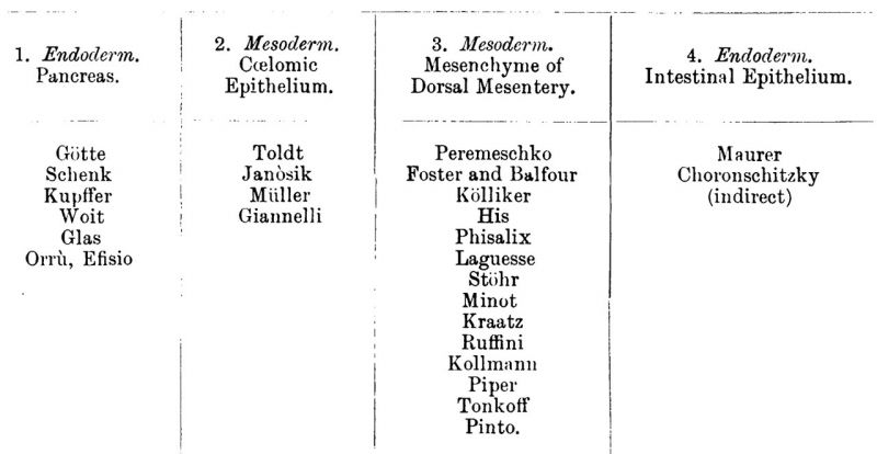 . I ' 1 l 1. Endotlemn. 1 2' gfffzm‘ bfiésgfiififdgfllléf 4. Endoderm. . Pancreas. Epithelium. D Orsal Mezenieryo Intestinal Epithelium.
It will be seen from the above table that the majority of writers have taken the view that the spleen is developed from the mesenchyme, and it might seem unnecessary to go once more into the question, but it must be remembered that many of the authors quoted have only touched superficially on the question, and have dealt with the development of the spleen in different classes of vertebrates.
Tonkoff’s important work deals only with the Ainniota, and, in spite of Pinto’s comprehensive and careful study of the question in Amphibia, Reptiles, Birds and Mammals, there still remains the fact that Maurer, and to some extent Choronschitzky, hold views which are widely different from those of other authors. It seemed worth while then, as the question, for Amphihia at any rate, is still open, to try and arrive at definite conclusions, and I therefore obtained a series of enibryos of Rana temp07'a'ria..
In the following account the most important details of the various stages are described.
Section II. Descriptive
My series of specimens include embryos of all stages from 10 mm. total length (4 mm. ano—buccal) to 27 mm. total length (11 mm. ano-buccal). (In older stages, the total length is no guide to the age of the embryo, and the ano-buccal length is not always dependable when the development of the limbs has begun.) I made both longitudinal and transverse sections, but found the latter the 111ore useful. Most of them were stained in bulk with haematoxylin and eosin.
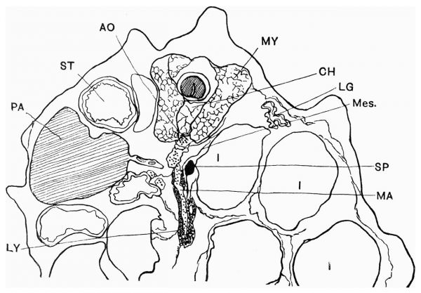
In an embryo of 27 mm. total length, in which the limbs have not yet appeared externally, the spleen appears as an oval body of smooth outline and of considerable size. It lies partly anterior to and partly on the left side of the mesenteric artery, in close contact with its wall, and appears in transverse sections as if wedged between the artery and the intestine which lies on its left, and to which it is attached by mesentery (see fig. 1). It is composed of definite lymphoid tissue and contains blood-spaces, but is very uniform in its appearance. It is quite clearlyseparated from the artery and the intestine, and lies in the mesentery surrounded by a distinct limiting membrane.
The spleen is comparatively late in developing and its growth is slow. Even when the limbs are well developed, the spleen shows very little advance on the condition to be seen in an embryo of 18 mm. total length.
Fig. 1. Transverse section of embryo of 18 mm. total length, showing position of spleen in relation to other organs. Ventral part of section omitted.
AO, aorta; CH, notochord; I, intestine; LG, lung; LY, lymphoid cells; MA,mesenteric artery; Mes., mesentery; MY, myotome ; PA, pancreas; SP, spleen; ST, stomach.
Stage I
(fig. 2). Embryo 10 mm. total lengtl1, 4 mm. ano-buccal. Stained methyl blue. Transverse sections of 10,u. Liver and pancreas well developed. Walls of intestine thick and convoluted. Many granulated cells containing yolk still to be found in them.
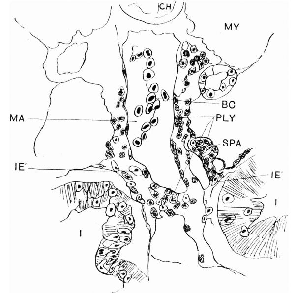
Mesenteric artery well developed, sending branches to pancreas and alimentary canal. The walls of the mesenteric artery are surrounded throughout by a thick coat of round cells with large nuclei (PLY). These are to be seen both on the branches and on the main trunk, and already a small heaping up of such cells may be seen on the left of the mesenteric artery, exactly in the position which the spleen will later on occupy (SPA).
The cells, although not unlike the cells of the intestinal epithelium, are not identical with them. Mitosis is certainly going on in the intestinal epithelium, but only a few of the epithelial cells show any indication of being squeezed out into the surrounding mesoderm (IE', fig. 2).
Fig. 2. Transverse section of embryo of 10 mm. total length, showing mesenteric artery with surrounding coat of lymphoid cells and the first indication of the spleen anlage. High power.
BC, blood-corpuscles; CH, notochord ; I, intestine ; IE’, strayed endodermal cells; MA, mesenteric artery; MY, myot--me; PLY, primitive lymphoid cells; SPA, spleen anlage.
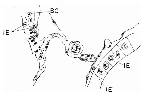
That this epithelial extrusion is not limited to early stages, nor to that part of the alimentary canal in the neighbourhood of the spleen, is shown in fig. 5, which shows such extrusion taking place. The section was taken through a part of the canal remote from the spleen, and at a stage when the spleen was already a definite" structure, covered by a limiting membrane.
In embryos of 10 mm total length, Where the spleen is in process of formation, the intestinal Walls are not unistratified as in Maurer’s figure, but are thick and convoluted, which makes it extremely difficult to determine in what plane mitosis takes place (fig. 2, I).
The epithelium is clearly divided in all situations from the mesenchyme lying outside it, and the round nucleated cells which are to be seen in the mesoderm among the more spindle-shaped cells are smaller and more granulated than the epithelial cells of the intestine, and . resemble more closely the cells surrounding the Walls of the arteries (fig. 2, PLY). Again, many'of these last-mentioned cells are undergoing mitotic division, although Maurer states that no such division could be seen in the connectivetissue cells at a similar stage. They are certainly “forerunners” of lymphoid cells, and are, even at this stage, grouped as thickly around the mesenteric artery itself as round its terminal branches, even although this is an earlier stage than any described by Maurer, and there is no indication that these cells are descendants of migrants from the intestinal epithelium.
It may be objected that the cells above mentioned are not definitely lymphoid in character; but the condition I have described, and which is shown in fig. 2, persists, and the spleen is developed by proliferation of the cells, which gradually become more lymphoid in character, and surround the dorsal part of the artery at the level at which the organ is situated when it is completely established. Exactly the same kind of cells are found, as Maurer himself says, in the connective tissue of the kidney, the genital gland, etc., which could not have travelled by the same path as those followed, according to him, by the spleen cells. I quote in this connection from Pinto, who says (xxvi. p. 388), “ Quello che risalta subito e il fatto che essa si presenta circondata come da una manicotto di grosse cellule rotonde con nuclei pure grossi, cellule che vanno diradandosi mano mano che andiamo verso le diramazioni arteriose che si sperdono nel mesenterio. Simili cellule troviamo anche qua e la sparse nel mesenterio ed alcune anche proprio al di sotto dell’ epitelio dell’ intestino.”
In other embryos of 10 mm. total length the thickening of the mesenchyme cells is more general, and no definite spleen anlage can be distinguished.
Stage II
Embryo 15 mm, stained methyl blue. Walls of intestine thinner and more simple. Single-layered epithelium shows distinct rounded body on the left side of the mesenteric artery. It is attached by mesentery to the part of the intestine lying to the left of it, and lies close against the wall of the artery. It extends through seven sections of 5,u, and is then lost as a lymphoid thickening on the wall of the mesenteric artery. It is covered by a single layer of coelomic epithelium, which is continuous With that refiected over the intestine and the body Wall. It is in very close contact with the arterial Wall, but is distinct from it. The cells are for the most part rounded, granular, with darkly stained round nuclei. They are in process of cell division, which can be seen in various stages. The tissue also contains blood corpuscles. There are no blood-spaces.
In some embryos even of slightly greater length the spleen is less definite. The lymphoid tissue is present in large quantity, and is heaped up around the artery at the level Where the spleen eventually comes to lie. In one series of sagittal sections the splenic thickness is very. Well marked, and the mesenteric artery passes through its posterior part. This is probably due to the fact that the spleen is closely associated with the lymphoid tissue surrounding the wall of the artery, and occasionally extends backwards into closer connection With it. The same remarks concerning the character of the cells apply as in the last stage described.
Scattered cells are to be seen which appear to be identical with the endodermal epithelial cells, and may have been extruded in the process of cell ‘division, but they are quite casually distributed, and the difference between them and the lymphoid cells is clearly marked (fig. 5).
Embryo 18 mm total length. Stained haematoxylin and eosin. The spleen runs through nine sections of .10“. It is considerably more definite than in the last stage, and lies freely in the mesentery, though in very close contact with the wall of the artery, which is still surrounded by scattered lymphoid cells, and with the part of the intestine which lies to its right. The coelomic epithelium covers the free surface, but there is not adefinite capsule (fig. 1, SP).
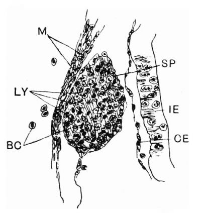
The cells composing it are already definitely lymphoid in character (fig. 3, LY). As compared with those of the intestinal epithelium, they differ markedly from the latter, being smaller, more irregular, and inclined to be elongated in shape and more granular, the. protoplasm taking a deeper stain, While the nucleus is more diffuse. At this stage mitosis is still proceeding in the cells of the intestinal epithelium, but it is diflicult even yet to determine the plane of division on which Maurer lays so much stress (fig. 3, IE). Here and there, cells derived from these mitotic divisions lie outside the basement membrane in the mesenchyme surrounding the intestinal wall. They are also to be seen here and there along the walls of branches of the mesenteric artery, but they do not collect specially round its terminal branches, as Maurer asserts, and they appear to be quite distinct from the lymphoid cells on the arterial Wall, which are to be seen in the same sections. A certain amount of emigration from the intestinal epithelium does take place, but there seems no reason for believing that the cells which have migrated take any direct part in the formation of the spleen.
Stage III
Embryo 24 mm. Sagittal section. Stained haematoxylin and eosin. The spleen runs through eighteen sections of 10,u.; it lies against the wall of the mesenteric artery, to the left side of and anterior to it. It is surrounded by a capsule of flattened epithelium, and is separated from the artery wall. It is dorsal and posterior to the stomach.
The cells are granulated, the majority are rather large and rounded, and the epithelial capsule is incomplete. Various stages of cell division can be seen in the splenic cells, many blood corpuscles are scattered through the tissue, and some dark elongated cells are present, which often divide transversely.
Fig. 3. Spleen in embryo of 18 mm. total length, lying between wall of mesenteric artery and wall of intestine. High power. Lymphoid cells of the spleen in various stages of division.
BC, blood-corpuscles ; CE, coelomic epithelium; IE, intestinal epithelium; LY, lymphoid cells; M, primitive muscle fibres; SP, spleen.
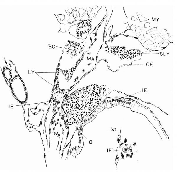
Embryo 27 mm. total length, 11 mm. ano-buccal. Stained haematoxylin and eosin (fig. 4). Shows the spleen as an oval, smooth body, running through thirty—five sections of 10”. It lies freely in the dorsal mesentery, by the side of the mesenteric artery. At its greatest diameter it is in very close contact with the artery, and with the intestine to the left of it, but it is separated from it, both by the coelomic epithelium and by its own distinct limiting membrane. In some sections a small blood-vessel is seen running between the intestine and the spleen, and sending branches into it. The dorsal mesentery is very diffuse and thin, and shows many sections through blood-vessels. Cells consist of (1) large cells with granular nuclei in various stages of cell division ; some are rounded, others spindleshaped or oval. (2) Smaller elongated cells, very deeply stained; these are possibly blood corpuscles in an imperfect state. (3) Red blood corpuscles (see fig. 4). In some sections small vessels can actually be seen entering the spleen tissue. There are no definite Malpighian corpuscles, but the Whole tissue is permeated with blood corpuscles.
Fig. 4. Transverse section of embryo of 27 mm. total length (ll mm. anobuccal), showing spleen, mesenteric artery with lymphoid cells around its walls, and intestinal epithelium. (a) A small portion‘ of mesentery near the arterial Wall, showing a wandered cell from the intestinal epithelium among the mesenchyme cells.
BC, blood-corpuscles; CE, coelomic epithelium ; IE, intestinal epithelium; IE’, strayed endodermal cells; LY, lymphoid cells; MA, mesenteric artery; MY, myotome; SLY, subchordal lymphoid tissue.
In an embryo in which the limbs were Well developed and the tail partly absorbed, the spleen shows Very little further advance in structure, but the capsule -is rather more developed. It Will be seen from fig. 4.« that the lymphoid tissue which surrounds the artery is less dense than in earlier stages of development, and is more conspicuous around the main trunk of the artery than at its terminal branches. The lymphoid mesenchyme cells (LY, fig. 4«) around the artery are exactly similar to those in the spleen itself, and differ markedly from the cells which appear to be. strayed endodermal cells from the epithelium of the alimentary canal.
In fig. 4 there are a few of these cells (IE’) to be seen lying in the mesentery among the mesenchyme cells. It will be seen that they differ in appearance from the latter, While they bear a strong resemblance to the cells of the intestinal epithelium, being large, clear, and spherical in shape, with very definite nuclei.
Fig. 5. From an embryo of 15 mm total length. Portion of intestinal wall, with small terminal branch of mesenteric artery and mesentery. White and red blood-corpuscles, mesenchyme cells in the mesentery, and strayed endodermal cells. One cell from the intestinal epithelium lies between it and the epithelium of the mesentery.
BC, blood-corpuscles ; IE, intestinal epithelium; IE’, strayed endodermal cells.
In the same series of sections, similar cells are to be seen lying in the mesentery in parts of the alimentary canal remote from the spleen, and they appear to be quite impartially, though sparsely, distributed.
The first possible forerunners of the lymphoid cells of the spleen are then to be found in embryos of 10 mm. total length (about 4 mm. anobuccal). The mesenteric artery at this stage has a thick coat of mesenchyme cells, which have begun to be heaped up in the position to be occupied by the spleen. These cells are grouped thickly round the main trunk and branches of the mesenteric artery, and in no instance did I find such cells surrounding the terminal branches, and not the artery itself. These cells differed in appearance from those of the intestinal epithelium in size, shape, and appearance of nucleus. Cells identical With the intestinal epithelium were to be seen here and there in the mesentery, but were few in number, and easily to be‘ distinguished from the mesenchyme cells among which they lay (fig. 4:, (ct) ). In no case were there such cells in the spleen anlage itself, nor was there any transition stage which could lead me to suppose that they were altering their character. The appearance of the above mentioned cells, both in the mesentery and in the mesenchyme surrounding the intestine, confirm Maurer’s View that there is a migration of intestinal epithelium, but the migration takes place along the whole length of the intestinal canal, and continues long after the spleen is a distinct structure and the cells take no part in spleen formation.
The cells were more frequent in the later stages than in the less developed embryos, and even at the stage at which Maurer first notes their appearance the spleen anlage has begun to appear.
Even before there is any trace of the actual spleen, the trunk of the mesenteric artery shows the thick coat of mesenchyme cells, which are undoubtedly primitive lymphoid tissue.
The coelomic epithelium certainly forms the capsule, but it does not appear to take a larger share than this in the development of the spleen. The limiting membrane is very distinct from the rest of the tissue throughout its development. (See Laguesse, X. p. 476.)
Conclusions
In the first place, it may be stated that the pancreas, in Rana at least, can have no part in the formation of the spleen. The two organs are not in contact at any stage in the development of either, and the pancreas is already well developed when the spleen “ anlage ” first appears.
Secondly, for reasons which I hope I have made clear in the last section, I do not believe that the endodermal epithelium of the intestine is responsible for the formation of the spleen, even indirectly. The migration of cells from the intestinal epithelium, where it does occur, takes place at a stage when the spleen “ anlage ” is already formed, and when the cells of which it is composed are themselves proliferating rapidly. However, that these epithelial cells do go to increase the mesodermal tissue of the 1nesentery seems clear, though I do not think the part they play is a very considerable one.
My conclusion then is, that the spleen in Rana arises from the mesenchyme tissue of the dorsal mesentery, in close connection with the mesenteric artery, as a thickening of the lymphoid tissue which surrounds the wall of the artery at an early stage. It develops by proliferation and differentiation of these primitive lymphoid cells, and becomes highly vascular. The coelomic epithelium appears to form the capsule, and perhaps enters into the formation of the reticular network.
In conclusion, I must express my gratitude to Professor Robinson for his most kind assistance and criticism, and for placing the necessary apparatus at my disposal While I was Working in the Anatomical Department at Birmingham University. My warmest thanks are also due to Dr Violet Coghill, whose constant help and suggestionhave been invaluable.
Bibliography
(1) PEREMESCHKO, “ Beitrag zur Anatomie der Milz und Leber und die Entwickelung der Milz,” Sitzungsberic/ate der Is. It. Akaclemie zu Wien, 1867.
(2) GOTTE, Beitrdge zur Entzoickelungsgeschichte des Darmkanals beim Hiihnchen, Tiibingen, 1867.
(3) MULLER, W., Handbuch der Lehre van den Geweben, Leipzig, 1871.
(4) SCHENK, Lehrlmeh der Embryol. «les Mensehen und der Wirbeltlziere, 1873.
(5) FOSTER and BALFOUR, Grundzage der Entwiclcelungsgeschielzte des Menschen und der Tiere, Leipzig, 1876.
(6) KOLLIKER, Entwickelungsgese/2ichte des Menschen und der Tiere, p. 579, 1879.
(7) His W. Anatomie Menschliche Embryonen I - Embryonen des ersten monats (Anatomy of human embryos - Embryos of the first month). (1880) Leipzig.
(8) PHISALIX, “Recherches sur Panatomie et la physiologic de la rate chez les Ichthyopsidés,” Archives «Ie Zool. expérimentale, 1885.
(9) TOLDT, C., “Zur Anat. der Milz,” Wiener klinische Wochensehmfl, 1889.
(10) LAGUESSE, E., Journal d’Anaz‘0mz'e et de la P/zysiologie, Paris, 1890; and “ La. Rate, est-elle d’origine mésodermique ou entodermique ‘E ” Bibliograplaie Anatomique, tome ii., 1894. A
(ll) MAURER, F., “Die erste Anlage der Milz und das erste Auftreten Von lymphatischen Zellen bei Amphibien,” Morphol. Jahrbuch, Bd. xvi.
(12) Kfiprmcn, C., “Ueber die Entwick. Von Milz und Pankreas,” Miinch. medicin. Abhandlungen, Miinchen, 1892.
(13) MINOT, Entzoickelungsgeschiclzte des Menschen, 1894.
(14) JANOSIK, “Le pancréas et la rate,” Bibliogr. Anat., iii., 1895.
(15) STGHR, “ Ueber die Entwickelung der Hypochorda und des dorsalen Pankreas bei Rana temporaria,” Morphol. Jahrbuch, Bd. xxiii., 1895.
(16) KBAATZ, “ Zur Entstehung der Milz,” Inaug. Diss., Marburg, 1897.
(17) Worr, 0., “Zur Entwickelung der Milz,” Anat. Hefte, 1897.
(18) RUFFINI, A., “Sullo sviluppo della Milza nella Rana esculenta,” Monitore Zoologico Italiano, x., 1899.
(19) CHORONSCHITZKY, B., “ Die Entstehung der Milz, Leber, Gallenblase, Bauchspeicheldriise und des Pfortadersystems bei den verschiedenen Abteilungen der Wirbelthiere,” Anat. I-Iefte, Bd. ix., 1899.
(20) KOLLMANN, J ., Le/zrbuch der Entwiclcelungsgeschichte des Mensclzen, Jena, 1 900.
(21) GLAS, “Ueber die Entwickelung der Milz bei Tropidonotus natrix,” Sz'tzungsber. der lcaiserl. A/cad. der Wissenschaften, Wien, 1900. Development of the Spleen 301
(22) GIAUNELLI, “A1cuni ricordi sullo "sviluppo della milza nei Rettili,” Am’ B. Accad. dei Fisiocritici, Siena, série iv., v., 12 anno, 209, 1900.
(23) TONKOFF, “Die Entwickelung der Milz bei den Amnioten,” Archie: fzlr mikcroskopischen Anatomie, Bd. lvi., 1900.
(24) ORRU, EFISIO, “Su11o sviluppo della milza,” Monitore Zoologico, 13 anno, 1902.
(25) PIPER, “Die Entwickelung von Leber, etc., bei Amia calva,” Arch. f. mils. Anat. u. Entw2'ckelungs., Supplement. Band, 1902.
(26) PINTO, “ Sullo sviluppo della milza,” Arclnivio Italiano dz’ Anatomia e Em briologia, 1904.
Cite this page: Hill, M.A. (2024, April 28) Embryology Paper - Development of the spleen. Retrieved from https://embryology.med.unsw.edu.au/embryology/index.php/Paper_-_Development_of_the_spleen
- © Dr Mark Hill 2024, UNSW Embryology ISBN: 978 0 7334 2609 4 - UNSW CRICOS Provider Code No. 00098G


