Paper - Data on the number of somites compared with age in the white rat
| Embryology - 27 Apr 2024 |
|---|
| Google Translate - select your language from the list shown below (this will open a new external page) |
|
العربية | català | 中文 | 中國傳統的 | français | Deutsche | עִברִית | हिंदी | bahasa Indonesia | italiano | 日本語 | 한국어 | မြန်မာ | Pilipino | Polskie | português | ਪੰਜਾਬੀ ਦੇ | Română | русский | Español | Swahili | Svensk | ไทย | Türkçe | اردو | ייִדיש | Tiếng Việt These external translations are automated and may not be accurate. (More? About Translations) |
Landacre FL. and Amstutz MM. Data on the number of somites compared with age in the white rat. (1929) Ohio J. Science. 29(6): 253-259.
| Online Editor |
|---|
| This historic 1929 paper by Landacre and Amstutz described development of the rat somites (somitogenesis). This paper includes a table (Table 3) summarising somitogenesis in the rat.
|
| Historic Disclaimer - information about historic embryology pages |
|---|
| Pages where the terms "Historic" (textbooks, papers, people, recommendations) appear on this site, and sections within pages where this disclaimer appears, indicate that the content and scientific understanding are specific to the time of publication. This means that while some scientific descriptions are still accurate, the terminology and interpretation of the developmental mechanisms reflect the understanding at the time of original publication and those of the preceding periods, these terms, interpretations and recommendations may not reflect our current scientific understanding. (More? Embryology History | Historic Embryology Papers) |
Data on the Number of Somites Compared with Age in the White Rat
F. L. Landacre And H. M. Amstutz
Department of Anatomy, University of California and Ohio State University.
Introduction
The following report containing data on the relation between age and the number of somites in the rat is based on materials secured from the rat colony of the Department of Anatomy at the University of California during the summers of 1925 and 1926.
The material was collected at the suggestion of Dr. Herbert M. Evans with the object of studying the whole problem of the origin of the neural tube and the relation of the neural crest and placodes to the origin of the cerebral ganglia.
The publication of an extensive paper by Adelman (’25)[1] on the neural folds and cranial ganglia of the rat, resulted in a change in the original program, the senior author being responsible for the later stages in development especially those involving the structure and possible contribution of the epibranchial placodes to the cerebral ganglia.
The effort to arrange a graded series of embryos, in order to follow the growth and differentiation of some particular structure which cannot be followed except in fixed material, always presents the uncertainty as to whether the series is as complete as possible and whether it is possible to predict the stage of growth and differentiation on some definite standard such as age or number of somites. When this can be done the grading of a series becomes a simple matter. When the variation between such criteria as age, length, weight and number of somites is pronounced then the problem of arranging a series to illustrate the growth of some particular body structure lacks the element of predictability largely, and the structure must be followed more or less independently and the uncertainty of stating that any particular phase of growth or differentiation is absent is very great.
In order to arrange embryos in a series that would be as nearly continuous as possible it was decided to base the series on the age of embryos estimated from the time of deposition of the sperm plug. The mating was done by Dr. Evans after the proper stage in the oestrus cycle had been determined from vaginal smears. Extreme care was exercised that the estimated time of deposition of the sperm plug should not vary in the records by more than five minutes although the age as recorded in the tables is given to the nearest hour. This was accomplished, by examining each female after coitus at intervals of not more than five minutes. After the deposition of the sperm plug as determined by vaginal examination the female was removed from the breeding cage.
All males used in the mating were tested for fertility prior to use for this series, but females had not been previously mated. The extreme care exercised in the choice of both parents as well as the accuracy of the time of deposition of the sperm plug seems to the writers to reduce to a minimum whatever objections there might be to using the deposition of the sperm plug as a means of estimating the age of embryos. Whatever defect this method may have, due to variation in the time of ovulation and fertilization as compared with the time of deposition of the sperm plug, it is a convenient and easily controlled method and gives a definite standard from which to make comparisons.
The use of females that had not been mated previously may be open to some objection since in the rodents at least the variations in growth and differentiation and possibly in age may vary more with first matings than with later matings.
A characteristic of the Berkeley colony should be noted although it is not apparent why it should effect the variations between age and number of somites. The colony is descended from a cross between an albino rat from the Wistar colony and a wild gray rat, Swezey (26-28).[2][3] The Berkeley colony shows two different chromosome counts, 42 and 62 with haploid numbers of 21 and 31 respectively, 42 being the number for both the albino and wild gray rat.
There is a further possibility that variations between the number of somites and age as estimated from the time of deposition of the sperm plug might be due to differences in time of fertilization and this variation might be greater in first matings than in subsequent matings.
The observations of Long and Evans ('22)[4] indicate that this difference as estimated by variations in ovulation is slight since ovulation occurs in first matings throughout a period of 12 hours, i.e., the last 12 hours of the II and III stages of the oestrus cycle, and following littering during a period of 8 hours, i.e., between 16 and 24 hours after littering, so that if one can estimate age by ovulation there should not be any marked variation in embryos due to first matings providing this variation is not greater between embryos of different mothers than between embryos of the two horns from the same uterus or between embryos from the same horn of the uterus. The four hours more or less assumed for the sperm to traverse the oviduct would normally transpire before ovulation was completed since the female accepts coitus during the II period of oestrus, i.e., presumably during the early part of the 30 hours involved in the II and III stages of the oestrus cycle and would not seem to furnish a basis for differences between age and number of somites. ‘ While the collection of material was going on, examination made on single litters under the dissecting microscopes showed such variations in size and number of somites that it became apparent that arranging a graded series based on age as estimated from the deposition of the sperm plug would present serious difficulties and that these same difficulties might be encountered in the effort to describe almost any phase of the formation of neural crest, ganglia, or placodes and their relation to each other.
In the paper by Adelman (’25)[1] cited above the conclusion was reached that there is no evidence for the contribution of cells from the epibranchial placodes to the ganglia of the VII, IX and X nerves in the White rat. Notwithstanding, the difficulties of following placodal cells as distinct from mesenchymal cells in birds and mammals, a difficulty which has been recognized for many years, there seemed to be a possibility for error in this case owing to difficulties of securing a series that was fairly continuous for any given structure when either age or number of somites was used as a criterion for determining where to look for a given step of the process if present.
Since marked variations occur not only between litters of the same age but between embryos of the same litter, both between embryos of the right horn as compared with the left and even within the same horn these variations even with the precautions mentioned above make it evident that any effort to predict what could be expected to occur in any given case based on age or number of somites might be hazardous to say the least.
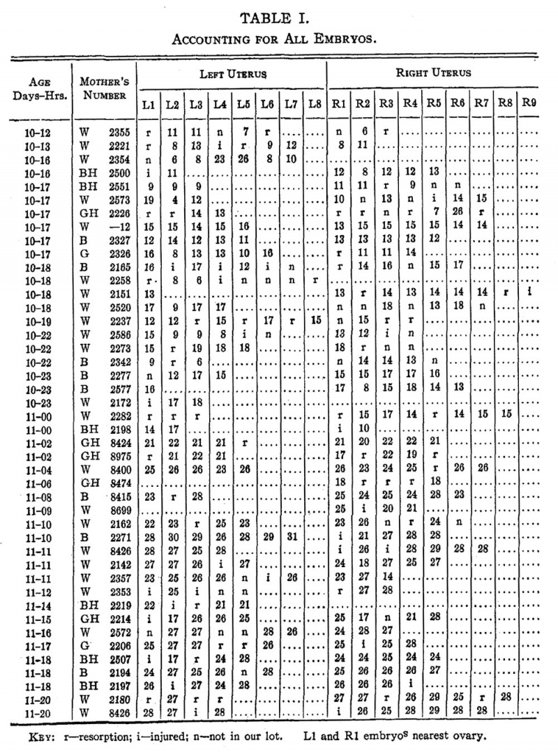
Table I. Accounting for All Embryos
KEY: t—resorption; i—injured: n--not in our lot. L1 and R1 embryos nearest ovary. As a. preliminary to the study of the variability in the formation of the placodes for instance as compared with age or number of somites the following tables are presented without any attempt to interpret them statistically except to call attention to their value as a basis for prediction since that is the practical value which such tables have when a series of embryos is arranged with a View to following any given structure in growth and differentiation.
Complete data are given in Table I showing the age to the nearest hour, the position of the embryo in the right or left horn of the uterus, the number of resorptions, the number of embryos injured so that counts of somites could not be made and the number of embryos not in our collection.
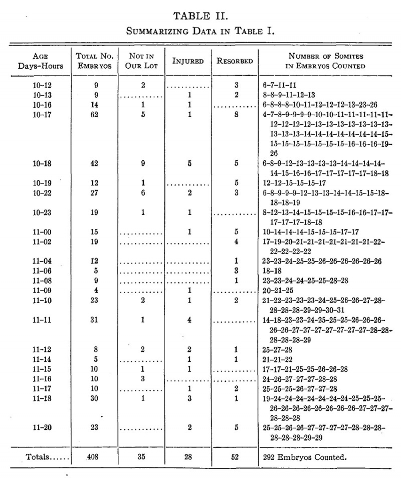
Table II. Summarizing Data In Table I
Table II is a summary of the data in Table I. A total of 408 embryos was found in the 44 litters. Of this number 35 embryos were sent to another university and no data are furnished in regard to them. They are marked “n ” in Chart I. Unfortunately 28 embryos were injured in excision or handling, so badly that the somites could not be counted. They are designated “i” in Chart I. fifty-two cases of definite resorption were found in the 408 cases observed and they are designated “r” in Chart I. finally an accurate count of somites in 292 embryos or about 75% of all uterine enlargements was secured. A very wide difference in number‘ of somites at many of the different ages should be noted. For example, at 10 days 17 hours the somite count ranges from 4 to 26, giving us an extreme variation. At 10 days 16 hours the range is from 6 to 26 somites, and similar variations are found at a number of other ages.
Table III. Showing Means and Variation for Each Age
| Maximum Variation | |||||||
|---|---|---|---|---|---|---|---|
| Age Days-Hrs. |
Number of Litters |
Number of Embryos Counted |
Means | All Embryos | One Litter | Standard Deviation |
Coefficient of Variation % |
| 10-12 | 1 | 4 | 8.7 | 6-11 | 6-11 | 2.8 | 32 |
| 10-13 | 1 | 6 1 | 0. 2 | 8-13 | 8-13 | 1.9 | 19 |
| 10-16 | 2 | 12 | 12.4 | 6-26 | 6-26 | 5.8 | 47 |
| 10-17 | 6 | 48 | 12.9 | 4-26 | 7-26 | 3 .3 | 26 |
| 10-18 | 4 | 23 | 14.1 | 6-18 | 9-18 | 3.1 | 22 |
| 10-19 | 1 | 6 | 14 .3 | 12-17 | 12-17 | 1.2 | 8 |
| 10-22 | 3 | 16 | 13.1 | 6-19 | 6-14 | 3.9 | 30 |
| 10-23 | 3 | 17 | 15.2 | 8-18 | 8-18 | 2.4 | 16 |
| 11-00 | 2 | 0 | 14.6 | 10-17 | 10-17 | 1.9 | 13 |
| 11-02 | 2 | 15 | 20 . 9 | 17-22 | 17-22 | 1. 3 | 6 |
| 11-04 | 1 | 10 | 25. 3 | 23-26 | 23-26 | 1.2 | 5 |
| 11-06 | 1 | 2 | 18.0 | 18-18 | 18-18 | .0 | 0 |
| 11-08 | 1 | 8 | 25 0 | 23-28 | 23-28 | 1.9 | 8 |
| 11-09 | 1 | 3 | 22 .0 | 20-25 | 20-25 | 2 . 2 | 10 |
| 11-10 | 2 | 18 | 26.2 | 21-31 | 21-31 | 2.9 | 11 |
| 11-11 | 3 | 26 | 25.6 | 14-29 | 14-27 | 3.2 | 12 |
| 11-12 | 1 | 3 | 26. 7 | 25-28 | 25-28 | 1.2 | 4 |
| 11-14 | 1 | 3 | 21.3 | 21-22 | 21-22 | .6 | 2 |
| 11-15 | 1 | 8 | 23.1 | 17-28 | 17-28 | 4 . 0 | 17 |
| 11-16 | 1 | 7 | 26.7 | 24-28 | 24-28 | 1.3 | 5 |
| 11-17 | 1 | 7 | 26.1 | 25-28 | 25-28 | 1.1 | 4 |
| 11-18 | 3 | 25 | 25 .4 | 19-28 | 19-28 | 1.9 | 7 |
| 11-20 | 2 | 16 | 27.2 | 25-29 | 25-29 | 1.2 | 4 |
| 23 Ages | 44 | 292 | Av 2.2 | Av 13% | |||
| Data Reference[5] Links: rat | somitogenesis | |||||||
| Table III Showing Means And Variation For Each Age | ||||||||||||||||||||||||||||||||||||||||||||||||||||||||||||||||||||||||||||||||||||||||||||||||||||||||||||||||||||||||||||||||||||||||||||||||||||||||||||||||||||||||||||||||||||||||||||||||||||||||||||||||||||||||
|---|---|---|---|---|---|---|---|---|---|---|---|---|---|---|---|---|---|---|---|---|---|---|---|---|---|---|---|---|---|---|---|---|---|---|---|---|---|---|---|---|---|---|---|---|---|---|---|---|---|---|---|---|---|---|---|---|---|---|---|---|---|---|---|---|---|---|---|---|---|---|---|---|---|---|---|---|---|---|---|---|---|---|---|---|---|---|---|---|---|---|---|---|---|---|---|---|---|---|---|---|---|---|---|---|---|---|---|---|---|---|---|---|---|---|---|---|---|---|---|---|---|---|---|---|---|---|---|---|---|---|---|---|---|---|---|---|---|---|---|---|---|---|---|---|---|---|---|---|---|---|---|---|---|---|---|---|---|---|---|---|---|---|---|---|---|---|---|---|---|---|---|---|---|---|---|---|---|---|---|---|---|---|---|---|---|---|---|---|---|---|---|---|---|---|---|---|---|---|---|---|---|---|---|---|---|---|---|---|---|---|---|---|---|---|---|---|
| ||||||||||||||||||||||||||||||||||||||||||||||||||||||||||||||||||||||||||||||||||||||||||||||||||||||||||||||||||||||||||||||||||||||||||||||||||||||||||||||||||||||||||||||||||||||||||||||||||||||||||||||||||||||||
| Reference: Landacre FL. and Amstutz MM. Data on the number of somites compared with age in the white rat. (1929) Ohio J. Science. 29(6): 253-259. | ||||||||||||||||||||||||||||||||||||||||||||||||||||||||||||||||||||||||||||||||||||||||||||||||||||||||||||||||||||||||||||||||||||||||||||||||||||||||||||||||||||||||||||||||||||||||||||||||||||||||||||||||||||||||
In Table III the arithmetic mean number of somites for all embryos of each age is given. It will be noticed that these means would form quite an irregular curve. It is probable that the means from many thousands of cases would form a much more gradual curve and show a definite gradual growth. However, there are still wide variations within the group at any one particular age.
The standard deviations for each age have been computed and are shown in Chart III. These range from 0 at 11 days 6 hours, at which age we have only two embryos, each having a count of 18 somites, to 5.8 at 10 days 16 hours. The significance of this standard deviation of 5.8 can be appreciated much more readily if the coefficient of variation is computed. We find that this gives us the enormous coefficient of 47%. Although that represents our extreme Variation for any one age, nevertheless, we have several other large coefficients and an average coefficient of 13%. This means that on the average each embryo contains a number of somites approximately 13% more or less than the mean for its age.
If we are liable to get coefficients of variation up to 47% for one particular age and can count on at least a deviation from the mean of 13% which would mean 2 or 3 somites and might mean as many as 12 or 13 somites, it seems reasonable to conclude that one is hardly justified in assigning a certain number of somites to an embryo of a certain known age. Further, in View of the fact that we have variations within a single litter of 7 to 26 somites (age 10 days 17 hours), it appears that one cannot be even reasonably sure of the number of somites in an embryo of any known age. The further inference is suggested that in the growth and differentiation of a particular structure when compared with a standard such as age, length, weight or number of somites in the rat, a wide range of variation might be encountered and might increase the difficulty of securing a complete series of growth changes.
Literature Cited
- ↑ 1.0 1.1 Adelman HB. The development of the neural folds and cranial ganglia of the rat. (1925) J. Comp. Neurol. 39(1): 19-171.
- ↑ Swezey O. The chromosomes of the rat. (1926) Science, N. S., 66:
- ↑ Swezey O. On the existence of two chromosome numbers in a mixed rat strain. (1928) J. Exp. Zool. 51(2):
- ↑ Long JA. and Evans HM. The oestrus cycle in the rat and its associated phenomena. (1922) Memoirs of Univ. of California. 6:
- ↑ 5.0 5.1 Landacre FL. and Amstutz MM. Data on the number of somites compared with age in the white rat. (1929) Ohio J. Science. 29(6): 253-259.
Online Editor - See also Painter TS. A comparison of the chromosomes of the rat and mouse with reference to the question of chromosome homology in mammals. (1928) Genetics. 13(2):180-189. PMID 17246549
Cite this page: Hill, M.A. (2024, April 27) Embryology Paper - Data on the number of somites compared with age in the white rat. Retrieved from https://embryology.med.unsw.edu.au/embryology/index.php/Paper_-_Data_on_the_number_of_somites_compared_with_age_in_the_white_rat
- © Dr Mark Hill 2024, UNSW Embryology ISBN: 978 0 7334 2609 4 - UNSW CRICOS Provider Code No. 00098G


