Paper - 1906 Observations on the Labyrinth of Certain Animals
| Embryology - 27 Apr 2024 |
|---|
| Google Translate - select your language from the list shown below (this will open a new external page) |
|
العربية | català | 中文 | 中國傳統的 | français | Deutsche | עִברִית | हिंदी | bahasa Indonesia | italiano | 日本語 | 한국어 | မြန်မာ | Pilipino | Polskie | português | ਪੰਜਾਬੀ ਦੇ | Română | русский | Español | Swahili | Svensk | ไทย | Türkçe | اردو | ייִדיש | Tiếng Việt These external translations are automated and may not be accurate. (More? About Translations) |
Gray AA. Observations on the labyrinth of certain animals. (1906) Proc. Royal Society of London. Series B. 78(525):284–296.
| Historic Disclaimer - information about historic embryology pages |
|---|
| Pages where the terms "Historic" (textbooks, papers, people, recommendations) appear on this site, and sections within pages where this disclaimer appears, indicate that the content and scientific understanding are specific to the time of publication. This means that while some scientific descriptions are still accurate, the terminology and interpretation of the developmental mechanisms reflect the understanding at the time of original publication and those of the preceding periods, these terms, interpretations and recommendations may not reflect our current scientific understanding. (More? Embryology History | Historic Embryology Papers) |
Observations on the Labyrinth of Certain Animals
By Albert A. Gray, M.D., F.B.S.E., Aural Surgeon to the Victoria Infirmary, Glasgow.
(Communicated by Professor John G. McKendrick, M.D., F.K.S. Received January 26, — Read February 15, 1906.) Plates 16—18.
- Links: Inner Ear Development | Embryology History
The method of preparing the membranous labyrinth devised by the writer[1] has made the study of that organ more easy. The difficulty which previously attended the examination of the inner ear was so great that even the extraordinary patience of Retzius only permitted him to complete the investigation of 5 mammals and 11 birds. In more recent times Alexander has examined several more by the microscope, but the total number altogether is still very small.
The writer has already published the results of his investigations in the seal and in man. The results of the examination of 14 other mammals are at present in course of publication and will not be described in this paper, except in so far as they throw light upon the subjects immediately under discussion. It is necessary, however, to refer in general terms to the anatomy of the inner ear in the case of the animals mentioned, in order that that of those described in this paper may be properly understood.
From an examination of 16 mammals the writer has found that the different orders and species present differences in the anatomy of the inner ear of three main types. These are (1) differences in the shape of the cochlea ; (2) differences in the size of the perilymphatic space of the semi- circular canals ; (3) differences in size of the otoliths.
The cochlea appears in the mammals under two types in addition to the peculiar type of the organ as found in the monotremes. These types are the sharp-pointed cochlea which is found in the carnivora and the rodents, and the flattened cochlea which is found in man, the monkeys, the lemurs, the ungulates and the cetacea. In the case of the carnivora one exception was found to this rule, the exception being the seal. This animal possesses a cochlea which is rather bowl-shaped than pointed like those of the cat, dog, and puma. There were no exceptions in the case of the ungulates, the least flattened cochlea being possessed by the pig, but the difference between it and the same organ in the other ungulates was found to be slight. There were no exceptions to the rule in the rodents : the rat, the mouse, the guinea-pig and the rabbit all being possessed of sharp-pointed cochleae. Three monkeys were examined and no exception was found, the cochlea being in every case of the flattened type and very like that of man, but smaller. The organ in the lemur was also of the flattened type, as was that of the porpoise.
The perilymphatic space of the semicircular canals proved to be a very interesting study. Our ideas of the anatomy of the labyrinth have depended so much upon the investigation of the organ in the human subject that it is not surprising that errors have crept in by assuming that certain features found in him will also be found in the lower animals. The present is a case in point. The perilymphatic space in the canals of the human subject is large and well developed, and it has been assumed that this would be true of other animals. But such is not the case. It so happens that man is one of the exceptions to a general rule. In mammals the rule is that the peri- lymphatic space is either very small or even completely absent in the canals. The exceptions to this rule are : — man, the monkeys, and the seal. No doubt there are other exceptions ; indeed, another falls to be recorded in this paper, but a sufficient number of examples have been examined to assert that the rule above given is a fairly general one.
In spite of the existence of this general rule, however, it is probable that the original type of the mammalian labyrinth possessed a well-developed perilymphatic space in the canals. The chief reason for this belief is, that in the reptiles and birds this is the type invariably found in all the animals of those divisions which have been examined. A more definite statement will be possible when the. labyrinth of the monotremes has been investigated in regard to this feature. One of these is at present in course of preparation, and the results of the examination will be published later. At present we may regard the presence of a well-developed perilymphatic space in the canals as indicating a labyrinth of a more ancient type. This, of course, does not mean that the particular species which possesses such a labyrinth is there- fore an ancient species. Obviously, man is a very recent species of animal, although he possesses the space referred to above. It merely means that man's progenitors have not dispensed with the space when other species of mammals were in process of losing it.
The presence or absence of this space is therefore a valuable guide to the relationship of different orders and species of mammals. Its value is enhanced by the fact that the space does not appear to have any physiological function. It is found in animals which have great delicacy of movement, and in animals which have not, as is evidenced by the monkeys on the one hand and by the sloth on the other. It is found in animals which migrate, such as the seal, and is absent in others which also migrate, such as the porpoise. In so far as the function of equilibration is concerned, the space appears to be of no value, for it is found in the climbing monkey, and is absent in the lemurs, which also are nearly all arboreal. In short, the space may apparently be retained or dispensed with according to the necessities of the case. What these necessities are we do not yet know, but it is very probable that if more room is required for the surrounding structures the perilymphatic space of the canals will disappear.
The otoliths of mammals have hardly in previous times been examined carefully, it being assumed that they are small, a natural assumption from the fact that in the human subject they are of very trivial dimensions. This assumption is in the main correct, but the writer has already discovered two exceptions, the porpoise and the seal, and another falls to be recorded in this paper, the kangaroo.
The labyrinths which are described in this paper are : the lion, the Indian gazelle, the three-toed sloth, and the kangaroo among mammals ; the crested screamer and the ostrich among the birds.
- ↑ Journal of Anat. and Physiology, 5 vol. 37, p. 379.
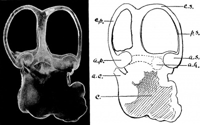
The Labyrinth of the Lion
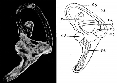
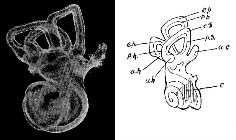
(Felis leo) Plate 16, fig. 1.
The membranous labyrinth of the lion differs in hardly any respect from that of other felidae except in the matter of size. The cochlea is of the sharp-pointed type, and measures 9 mm. in diameter in the lowest whorl. The second whorl measures 6 mm. in diameter. The scala tympani shows a marked bulging at its lower extremity just before it reaches the round window. This is a common feature in the carnivora and in some of the other mammals. The slant height of the cochlea from the upper margin of the round window to the apex of the organ is 4*5 mm. in length. There are three turns in the spiral of the cochlea, this being a fraction of a turn more than that of the puma, and a quarter of a turn less than that of the dog. The cat, like the lion, has three turns in its cochlea.
The vestibule measures 5 mm. in its longest diameter, and there are no otoliths in the cavity of a size sufficient to be recognised by the naked eye. The oval window is elliptical in shape, being in this respect like those of the other carnivora, except the seal, in which the aperture is semicircular (if the contradiction in terms may be excused).
The semicircular canals are very regular in shape, rounded, and without any noticeable irregularities such as are found in some mammals. Each canal lies in one plane, there being no lateral deviations. The superior is the largest of the canals. It measures 6*5 mm. from limb to limb internally, and 7*5 mm. externally. The height of the vertex of the canal from the vestibule is 6 mm., and the diameter of the canal itself at the vertex is 0*75 mm.
The posterior canal lies in a plane almost at right-angles to that of the superior canal. It is somewhat smaller than the latter. Its diameter, measured internally, is 5*5 mm. in length, and measured externally is rather less than 7*5 mm. The height of the vertex of the canal above the vestibule is 5*5 mm. and the diameter of the canal itself at the vertex is 0*5 mm.
Unfortunately the horizontal canal was broken in the process of preparation, but it was noticed that it was distinctly smaller than either of the other two. The plane of this canal is relatively low and, indeed, at its posterior extremity it appears to open into the ampulla of the posterior canal. This low level of the plane of the horizontal canal is very common among mammals, and in none of the mammals examined by the writer is the plane of the canal so high relative to the posterior canal as in the monkeys and in man. In the two latter the space enclosed by the curve of the posterior canal is almost bisected by the plane of the horizontal canal.
The perilymphatic space is almost entirely absent from the canals of the lion, indeed it can only be seen in the angles formed by the ampullae of the canals with the canals themselves. This feature is common to the carnivora that have been examined with the single exception of the seal. In the latter the space is well marked.
The length of the whole labyrinth of the lion is 17 mm. The cochlear portion is large relative to that portion formed by the vestibule and canals. In this respect the lion resembles the cat and other carnivora. A condition exactly the reverse of this is found in the lemur.
The angles which the planes of the canals form with one another are rather large in the lion. That is to say, the canals diverge widely from one another, much more so than in man. This divergence of the canals is greater in the lemur than in any animal which I have yet examined.
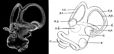
The Labyrinth of the Indian Gazelle
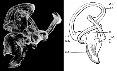
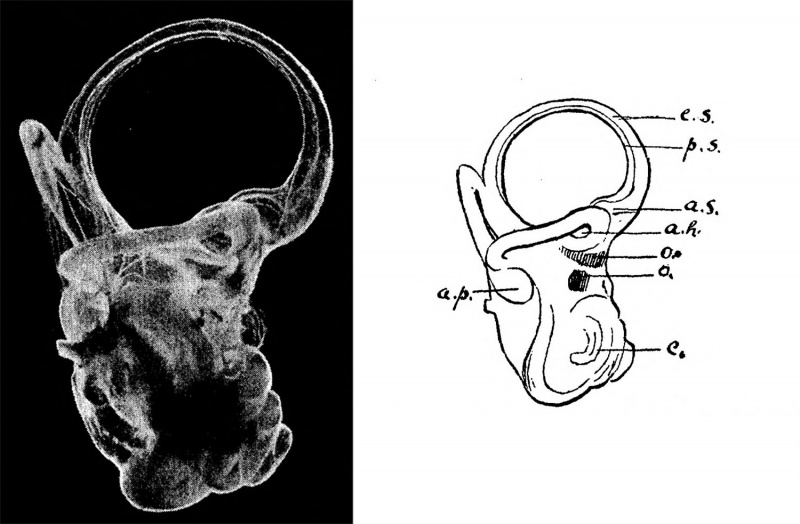
(Gazella Bennetti) Plate 16, fig. 2.
The membranous labyrinth of the Indian gazelle resembles that of the other ungulates in its general outline. There are, however, some unexpected differences.
The organ measures 14 mm. in extreme length from the outermost point on the vertex of the posterior canal to the innermost point on the lowest whorl of the cochlea. The diameter of the lowest whorl of the cochlea is 7 mm., while that of the second whorl is 3*5 mm. The diameter of the tube of the cochlea immediately in front of the round window is also 3*5 mm., the scala tympani showing a marked bulging downwards similar to that found in other ungulates and in most of the mammals. The slant height of the cochlea measured from the upper margin of the round window to the apex of the organ is 5*75 mm. The aqueduct of the cochlea is quite unlike that of any of the mammals which I have hitherto examined. Instead of being comparatively straight, as in its near ally the antelope, it is sharply curved. A large vein accompanies the aqueduct out of the cochlea and, to judge from the photograph, the blood is carried away from the cochlea by this vein. This disposition of the veins of the organ is different from that of the arteries, which are supplied by the internal auditory artery which enters the labyrinth by way of the internal auditory meatus.
The shape of the cochlea differs in no way from the general type of the ungulates, that is to say, it is of the flattened type. It consists of two and a-half turns.
The vestibule of the Indian gazelle is also like that of the other ungulates. It measures 3*75 mm. in its longest diameter and does not contain any otoliths of a size sufficient to be recognised by the naked eye. The oval window measures 2*75 mm. in its longest axis.
The semicircular canals of the Indian gazelle differ from those of the other ungulates in one important respect. The perilymphatic space is much more marked than in either the antelope, the sheep, or the pig. In the last- mentioned this space does exist throughout the whole length of the canals, but is so small that at parts it can hardly be seen ; in the sheep and antelope it cannot be seen at all in a large portion of the length of the canals. In the gazelle, however, the space can be traced easily round the whole course of all the canals. It is not so large as in man, the monkeys, the seal or the sloth. In their general appearance the canals show much the same features as those of the other ungulates, apart from the fact of the large perilymphatic space. They are regularly curved in outline, the pig being the one exception to this rule in the ungulates.
The diameter of the superior canal, measured internally, is 4*75 mm., while externally the diameter is 6 mm. The height of the vertex of the canal above the vestibule is 6 mm. and the diameter of the canal itself at the vertex is 1 mm. The posterior canal measures 4 mm. in its internal, and rather more than 6 mm. in its external diameter. The height of the vertex of the posterior canal above the vestibule is 5 mm. and the diameter of the canal itself at the vertex is 1 mm. The external canal measures 3 '5 mm. in its internal and 5 mm. in its external diameter. The height of the vertex of the canal above the vestibule is 3*5 mm. and the diameter of the canal itself at the vertex is 1*25 mm.
The canals of the Indian gazelle do not diverge from one another at quite such large angles as those of the antelope and the sheep. In this respect they resemble the canals of the pig, though the divergence is still less in the last-mentioned animal.
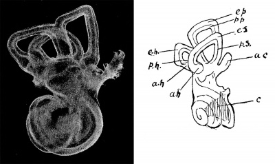
The Labyrinth of the Three-toed Sloth
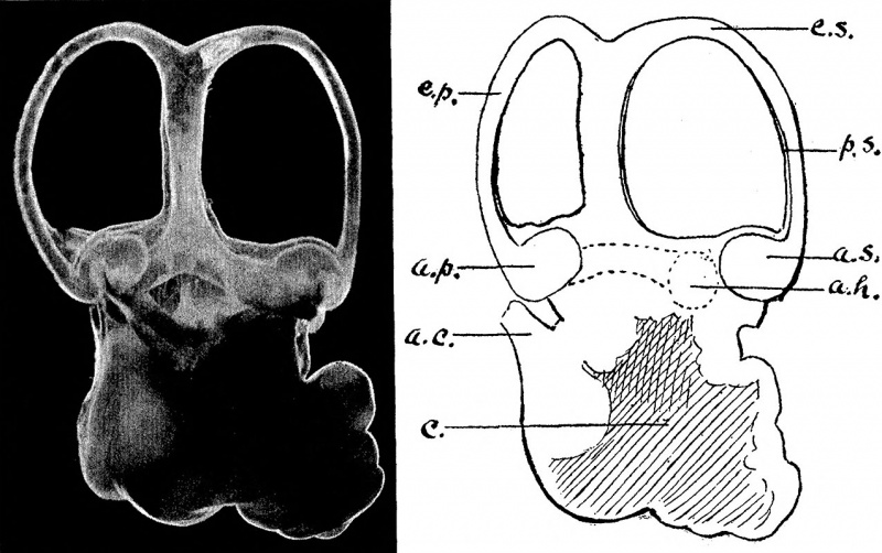
(Bradypus tridactylus) Plate 17, fig. 3.
Hitherto the writer has only had the fortune to obtain one example of the edentata, that being the three-toed sloth. To judge from this example, the labyrinth of these peculiar animals will be interesting and instructive to study.
The cochlea is intermediate in shape between the flattened and the sharp- pointed type, but inclining rather to the former. In this respect the organ differs from the great majority of mammals, since, as has been already pointed out, there is only one other animal among all those which have been examined which does not fall clearly into one of the two types, that animal being the seal.
The labyrinth measures 10 mm. in extreme length from the outermost point on the posterior canal to the innermost point on the lowest whorl of the cochlea.
The lowest whorl of the cochlea is 5*5 mm. and the second 3*75 mm. in diameter. The diameter of the tube of the cochlea in front of the round window is 2 mm. and there is no marked bulging of the floor of the scala tympani in this region. In the latter respect the labyrinth resembles that of man and the monkeys ; but the aqueduct of the cochlea of the sloth is much thicker than in these two orders. There are two and a-half turns in the cochlea and the slant height of the organ, measured from the upper margin of the round window to the apex, is 3*75 mm.
The longest axis of the oval window is rather less than 1*5 mm. in length, and the longest diameter of the vestibule is 3 mm. There are no otoliths of a size sufficient to be recognised by the naked eye.
The canals of the sloth are quite unlike those of any mammal which the writer has had the opportunity of examining. They are not semicircular in shape, the horizontal canal being the only one that approaches this form, and even it is irregular. The posterior and the superior canals are quadrilateral, or roughly so. The common limb of these two canals arises from each respectively at right angles, because there is none of that curving downwards to meet each other as they approach, such as is found in all other mammals which have been examined. Similarly, the canals as they approach their ampullary extremities do not curve towards the vestibule, but turn suddenly downwards at right angles a short distance before they dilate into the ampullae.
The sloth, in common with man, the monkeys, and the seal, has a well- developed perilymphatic space in all the semicircular canals. According to the view expressed by the writer this indicates a relatively ancient type of labyrinth.
The internal diameter of the superior canal measures 3 mm., and the external diameter 4*5 mm. The height of the vertex of the canal above the vestibule is 2*25 mm., and the diameter of the canal itself at the vertex is 1 mm. The internal diameter of the posterior canal is 2*5 mm., and the external diameter is 4*5 mm. The height of the vertex of the canal from the vestibule is 2*5 mm., and the diameter of the canal itself at the vertex is 1 mm. The external canal is much the smallest of the three, measuring only 1*5 mm. in diameter internally and 3*5 mm. externally. The height of the vertex of this canal above the vestibule is only 1*5 mm.,, and the diameter of the canal itself at the vertex is 1 mm.
The angles at which the canals diverge from one another are smaller than in any of the mammals which have been examined and this gives the canals the appearance of having been pressed somewhat together. In addition to this it will be seen, on comparing the measurements or on examining the photograph, that the vestibule and canals occupy a smaller proportion of the whole labyrinth than in most mammals. It is exactly the reverse, for example, of the condition found in the lemur, where the canals are very long and slender. It may be that this small size of the canals, associated with their irregular shape, may be in some way related to the sloth's clumsy and slow movements. The life which they lead, with the body inverted, as it almost continually is, may also be connected in some way with the curious development of these organs.
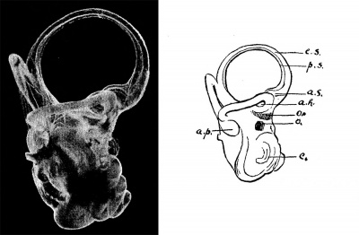
The Labyrinth of the Brush-tailed Wallaby
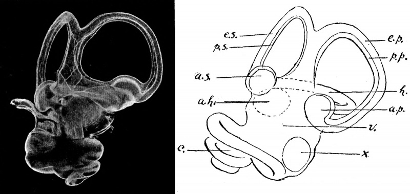
(Petrogale penicillata) (Plate 17, fig. 4.)
The labyrinth of the marsupials is not so divergent in structure from that of the general type of mammalian labyrinth as might be expected in an order of animals so far removed from most of the present divisions. It is, for example, less peculiar than that of the sloth and far less peculiar than that of the monotremata, the cetacea, or the seal.
The whole labyrinth measures 8 mm. in length from the outermost point on the vertex of the posterior canal to the innermost point on the lowest whorl of the cochlea.
The diameter of the lowest whorl of the cochlea is rather less than 4.5 mm. in length, while that of the second whorl is only 2.5 mm. in length. There is a marked bulging of the floor of the scala tympani in the region of the round window and the tube of the cochlea at this point measures 2*5 mm. in diameter. There are a little more than two and a-half turns in the cochlea and the slant height of the organ from the apex to the upper margin of the round window is 3.75 mm. The general shape of the cochlea is like that of the ungulates and more particularly like that of the pig. The aqueduct of the cochlea is a short tube of about 1.5 mm. in length, this being perhaps the most peculiar feature of the organ in this animal. The aqueduct is thick in proportion to its length and is straight. It is not triangular as in most of the mammalia, but is flattened from above downwards.
In proportion to the rest of the labyrinth the vestibule is rather large, measuring 4.0 mm. in its longest diameter. The longest axis of the oval window is 2*0 mm. in length. There are two otoliths of considerable size in the vestibule. They are larger than those found in any other mammal, with the single exception of the seal, even the porpoise not being an exception in this respect. Both otoliths lie on the inner wall of the cavity, one anteriorly immediately behind the ampullae of the superior and external canals, while the second lies a little below the first. They are both flat, and are of irregular outline.
The semicircular canals are very like those of other mammals and are beautifully regular in outline, reminding one of the same structures in the antelope, though of course much smaller. The superior canal measures 3*5 mm. in its internal, and 5 mm. in its external diameter. The height of the vertex of the canal above the vestibule is 3 mm., and the diameter of the canal itself at the vertex is 0*5 mm. The internal diameter of the posterior canal is 3 mm., and the external diameter of the same canal is 4.5 mm. The height of the vertex of the canal above the vestibule is 3 mm. and the diameter of the canal itself at the vertex is 0.5 mm. Thus the posterior canal is smaller than the superior. The smallest of the three canals is the external, which measures 3 mm. in its internal and 4 mm. in its external diameter. The height of the vertex of the canal above the vestibule is 2.75 mm., and the diameter of the canal itself at the vertex is 0*5 mm.
There is a very small perilymphatic space in the canals ; it is more noticeable in the angles formed by the ampullae with the canals themselves. In this respect therefore, the labyrinth is like that of the ungulates, with the exception of the gazelle.
The labyrinth of the kangaroo is not of such an ancient type as we might have expected, save in one respect — the presence of large otoliths. It is unfortunate that the writer has only been able to obtain one example of the marsupials, and it may be that in other species of this order a more ancient type of labyrinth may yet be found. In this connection it should be pointed out that the diprotodont class of marsupials is generally considered to be of more recent origin, and this, if true, may account for the fact that the labyrinth of the kangaroo is not of such an ancient type as we might have
expected.
The Labyrinth of Birds
It is quite outside the scope of this paper to describe in detail the typical labyrinth of birds. That work has already been done by several writers and in particular by Retzius, to whose writings the reader is referred. The present purpose is to show the likenesses and differences which exist between the various species. The reader need only be reminded that there are, as in almost all vertebrates, three canals which occupy nearly the same position relative to one another as they do in the mammals. On the whole, perhaps, they vary more in their disposition than do those of the mammals. One feature seems to be peculiar to the labyrinth of the bird : the horizontal canal, instead of terminating almost in the plane of the posterior canal, passes underneath the latter and projects backwards behind the plane in which it lies. The result of this arrangement is that the two canals form a cross of which the limbs are almost at right angles to one another. At the point of crossing there is a channel of communication between the two canals and it has been supposed that this is a constant feature of the labyrinth of the bird. It certainly is a very general condition, but, as will be seen later, it is not absolutely true of all the birds.
The perilymphatic space of the semicircular canals of the bird is always well marked so far as the present writer's investigations go. This is a notable distinction of the labyrinth of the bird from that of the mammals.
The shape of the canals in the bird is in general that of an ellipse rather than of a semicircle. The superior canal, however, varies considerably in shape, and no constant types can at present be described. This canal is always the largest, or rather has been found to be so, in all the examples hitherto examined.
The vestibule is relatively small. It usually contains otoliths of a size easily seen by the naked eye. They are flattened and are of various shapes. The most usual number of otoliths found in the vestibule is two. The cochlea of the bird consists of a more or less straight tube. It passes a little downwards and then inwards and forwards. There is usually a slight curve on it, with the concavity directed backwards and a little upwards. The main branch of the cochlear nerve runs along the posterior border of the cochlea and sends filaments forwards to the organ of Corti. At the tip of the cochlea, however, the nerve radiates out like a fan into the lagena, and at this spot there is in many birds a saddle-shaped otolith with the concavity directed outwards.
The ampullae of the semicircular canals of birds differ from those of the mammalia. They are usually set at a more acute angle with the canal as it leaves them. The nerve to the ampulla cuts into it, so to speak, and partially divides the ampulla into two portions, one adjacent to the vestibule and the other adjacent to the canal itself.
The Labyrinth of the Crested Screamer
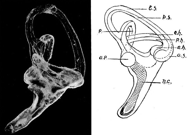
(Cariama cristata) Plate 18, fig, 5.
The term " crested screamer " is applied to two quite different birds. That from which the labyrinth was taken and prepared by the writer and forms the subject of this description, is closely allied to the cranes and has no relation to Ghauna cristata, which is related to the ducks and geese. According to some ornithologists, Cariama cristata is more closely allied to the hawks than to the cranes. The bird lives in the southern parts of Brazil and Paraguay. It will only fly if hard pressed, the usual method of progress being a stooping run. In some of its habits it is like a bustard, its note is a scream or bark. It lives in the high grass and the habits of the bird are diurnal.
The labyrinth is rather large for the size of the bird, measuring 15 mm. from the uppermost point on the vertex of the superior canal to the inner- most point at the tip of the cochlea.
The cochlea is very straight in this bird, the usual curve being almost entirely absent. It measures 6 mm. in length from the front of the round window to the tip of the organ. This is a long cochlea for a bird, that of the ostrich being the only one out of the nine which have been examined that is longer. The diameter of the tube of the cochlea immediately in front of the round window is 2 mm. in length. At the tip of the cochlea the nerve widens out into a spade-shaped structure, and a minute straight otolith is present at this point.
The vestibule measures 3 mm. in its longest diameter, and there is a very small otolith present in the utricle. The macula neglecta is to be seen close to the opening of the cochlea. The superior canal is much the largest and, roughly speaking, is in the form of an ellipse, with the long axis directed upwards and backwards.
The superior canal measures 6 mm. internally and 8 mm. externally. The height of the vertex of the canal above the vestibule is 6 mm. and the diameter of the canal itself at the vertex is 1 mm. The posterior canal is next in size to the superior and is also in the form of an ellipse. It measures 4 mm. in its internal diameter and 6*5 mm. externally. The height of the canal above the vestibule is 3 mm., and the diameter of the canal itself at the vertex is 1*5 mm. The common limb of the superior and posterior canals is very much shorter than is the case in any of the mammals. The horizontal canal is the smallest of the three and is elliptical in shape, the major axis being in the antero-posterior plane. It measures 3*5 mm., in internal and 6 mm. in external diameter. The height of the canal at the vertex from the vestibule is 3*25 mm. and the diameter of the canal itself at the vertex is 1*5 mm.
As is the case in most of the birds there is a communication between the horizontal and superior canals at the point at which they cross, but the opening is very small. The horizontal canal does not project so far back- wards in the crested screamer as in the majority of birds and this gives to these two canals an arrangement similar to that found in the mammals. The perilymphatic space is well marked in all the canals.
The Labyrinth of the Masai Ostrich
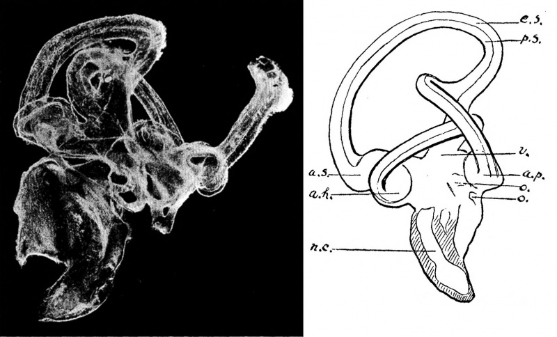
(Struthio masai). Plate 18, fig. 6.
In so far as the writer's investigations go, no examination has been made of the labyrinth of this division of the order of birds. It is, however, one of the most interesting on account of the fact that these birds are less distantly removed from the reptiles than any others.
Unfortunately both the labyrinths which the writer obtained were broken, and it is therefore impossible to give a description of the complete organ. The most important parts were not destroyed.
So far as can be judged from the broken specimens, the whole organ measures about 17 mm. in length. The cochlea is relatively small and only measures 6*5 mm. in length. It has a stumpy appearance and the backward curve is very well marked. The nerve spreads out fanlike at the tip of the organ and at this point there are several small otoliths arranged in the form of a saddle with the concavity directed outwards. This plurality of the otoliths at the apex of the cochlea is not found in any of the other birds which have been examined, though the single otolith which is so often found here is also saddle-shaped *and has the concavity in the same direction. The diameter of the tube of the cochlea just in front of the round window is rather more than 2 mm.
The vestibule is an irregular cavity and measures a little more than 3 mm. in its greatest diameter. It contains two large otoliths. Both of these are flat. The largest, which lies close to the ampullary openings of the superior and horizontal canals, is roughly circular in shape. The second and smaller one is almost square and lies about 1 mm. below and internal to the first. Both the otoliths are milk-white in colour. The oval window measures 2*5 mm. in its longest diameter.
As far as can be judged from the broken specimen, the superior canal is in the form of an ellipse, with the longest diameter lying backwards and upwards. It is much the largest of the three, measuring 7*5 mm. in its internal and 11 mm. in its external diameter. The height of the vertex of the canal above the vestibule is about 9*5 mm. and the diameter of the canal itself at the vertex is 1*5 mm. The posterior canal is also in the shape of an ellipse, with the long diameter in a vertical plane. It measures 5*5 mm. in its internal and 8*5 mm. in its external diameter. The height of the vertex of the canal above the vestibule is only 3*5 mm. and this gives to the canal a somewhat squat appearance. The diameter of the canal itself at the vertex is 1*5 mm. The horizontal canal is the smallest of the three and, like the posterior, is of a squat appearance. It measures rather more than 4 mm. in its internal and 8*5 mm. in external diameter. The diameter of the canal itself at the vertex is 1*25 mm.
A very interesting feature of the canals of the ostrich is the fact that there is no communication between the posterior and horizontal canals at the point at which the arch of the latter passes under that of the former. There is a distinct though narrow interval between them which is filled up with bone in the unprepared subject. This feature of the labyrinth of the ostrich is unique in birds so far as present investigations have shown, but it may be found in other birds of the same class. A specimen of the rhea is now in course of preparation and it will be interesting to see if this feature is repeated in that bird.
Explanation of Plates
Each labyrinth is represented by two plates. The upper plate is a half-tone reproduction from a photograph of the organ. The finer details are shown in this plate, but the three dimensions which the organ occupies are not of course appreciable and the comparative sizes are not represented, as the degree of magnification varies in each. The rough outline drawings below give, to a certain extent, the sense of three dimensions. The comparative sizes of the objects are also shown in these drawings, each being magnified about four times.
The lettering and magnification refer only to the outline drawings. The position of the object is not always the same in the drawing as in the photograph.
| The lettering is the same for all the plates. | ||
|---|---|---|
|
|
|
Fig. 1. Membranous Labyrinth of the Lion, Felis leo
Fig. 2. Membranous Labyrinth of the Indian Gazelle, Gazella Bennetti.
Fig. 3. Membranous Labyrinth of the Three-toed Sloth, Bradypus tridactylus.
Fig. 4. Membranous Labyrinth of the Brush-tailed Wallaby, Petrogale penicillata
Fig. 5. Membranous Labyrinth of the Crested Screamer, Cariama cristata
Fig. 6. Membranous Labyrinth of the Masai Ostrich, Struthio masai
| Historic Disclaimer - information about historic embryology pages |
|---|
| Pages where the terms "Historic" (textbooks, papers, people, recommendations) appear on this site, and sections within pages where this disclaimer appears, indicate that the content and scientific understanding are specific to the time of publication. This means that while some scientific descriptions are still accurate, the terminology and interpretation of the developmental mechanisms reflect the understanding at the time of original publication and those of the preceding periods, these terms, interpretations and recommendations may not reflect our current scientific understanding. (More? Embryology History | Historic Embryology Papers) |
Cite this page: Hill, M.A. (2024, April 27) Embryology Paper - 1906 Observations on the Labyrinth of Certain Animals. Retrieved from https://embryology.med.unsw.edu.au/embryology/index.php/Paper_-_1906_Observations_on_the_Labyrinth_of_Certain_Animals
- © Dr Mark Hill 2024, UNSW Embryology ISBN: 978 0 7334 2609 4 - UNSW CRICOS Provider Code No. 00098G

