Meyer - Essays on the History of Embryology 4
| Embryology - 28 Apr 2024 |
|---|
| Google Translate - select your language from the list shown below (this will open a new external page) |
|
العربية | català | 中文 | 中國傳統的 | français | Deutsche | עִברִית | हिंदी | bahasa Indonesia | italiano | 日本語 | 한국어 | မြန်မာ | Pilipino | Polskie | português | ਪੰਜਾਬੀ ਦੇ | Română | русский | Español | Swahili | Svensk | ไทย | Türkçe | اردو | ייִדיש | Tiếng Việt These external translations are automated and may not be accurate. (More? About Translations) |
Meyer AW. 1932 - Essays on the History of Embryology: Part I | Part II | Part III | Part IV | Part V | Part VI | Part VII | Part VIII | Part IX | Part X | Part XI | Arthur Meyer | Historic Embryology Papers
| Historic Disclaimer - information about historic embryology pages |
|---|
| Pages where the terms "Historic" (textbooks, papers, people, recommendations) appear on this site, and sections within pages where this disclaimer appears, indicate that the content and scientific understanding are specific to the time of publication. This means that while some scientific descriptions are still accurate, the terminology and interpretation of the developmental mechanisms reflect the understanding at the time of original publication and those of the preceding periods, these terms, interpretations and recommendations may not reflect our current scientific understanding. (More? Embryology History | Historic Embryology Papers) |
Essays on the History of Embryology IV
By A. W. Meyer, M. D.
Stanford University
- This is the fourth paper of a series of essays on this subject. Previous papers were printed in this journal as follows: Part I in December California. and Western Medicine, page 447; Part II in January California and Western Medicine, page 40; Part III in February California and Western Medicine, page 105.
SHORTLY before Malpighi’s observations on the development of the chick were published, an epoch-making observation had been made by a Dutch microscopist, Antoni Leeuwenhoek. This man, who was called an “immortal dilettante” by Professor Becking, a young countryman of his, is usually given credit for the discovery of the spermatozoon. Although Leeuwenhoek may have been a dilettante, he nevertheless made many important discoveries with microscopes made by himself, but which were far superior to any others of the time and were made by the hundred, according to Stein.
The Spermatozoon
It is regrettable that Leeuwenhoek’s imagination, like Swammerdam’s and Harvey’s, led him sadly astray. It seems he reported to the Royal Society, to which he had sent so much that was startling and what commissions could not confirm because they had inferior microscopes, that he was able to identify male and female spermatozoa by inspection alone!
Spermatozoa were first seen in 1675 by Hamm, a student of Leeuwenhoek, who is otherwise unknown, in the semen of a man “who had cohabited with an unhealthy woman.” It is to the credit of Leeuwenhoek that he quickly apprehended the significance of this discovery, and surmised that the moving, motile bodies which he called seminal filaments really were the male germs of animals. He looked for and found them in the testes of the dog and the rabbit, of birds, frogs, fish, and insects, and also in the tubes and uteri of dogs and rabbits. As Singer emphasized in “A Short History of Medicine,” this was a very deserving and important accomplishment in embryology, and he did other things, as Stein and others showed so well. Leeuwenhoek also estimated the total number of spermatozoa in the gonads of some animals and stated that those of codfish contained more than ten times as many sperm as there were inhabitants on the earth at that time. Since there are over a hundred million spermatozoa in a single cubic centimeter of semen of some mammals, and since he thought that the population of the earth might be over thirteen billions, it is evident that Leeuwenhoek approximated the ‘truth fairly well.
Animalcules had long been known to occur in printer’s ink, vinegar, and also in putrefying substances, hence it is easy to understand‘ that the presence of similar organisms in human semen not only aroused skepticism and evoked surprise, but also caused disgust. Cole quotes Andry as saying in 1701 that, “If, after you have taken off one testicle [from a dog] and by the aid of the Microscope examined the Humour that comes out of the deferent vessels, you shall discover in it such a hideous number of little worms that you shall hardly be able to believe your own Eyes.” But the indispensability of spermatozoa in procreation was not established experimentally until 1780, a century later, when Spallanzani performed the first experimental fertilization in toads, water salamanders and frogs, and proved that filtration of the semen destroys its fertilizing power and that an aura spermatica does not exist.
Whether Hartsoeker, who represented a flexed homunculus in the head of a spermatozoon, or Plantade, who signed himself Dalenpatius, and similarly represented a miniature human being in the extended posture, are to be taken seriously remains a question. Hartsoeker seems to have been known as unreliable and Plantade as a joker. Hartsoeker also stated that the tail of the spermatozoon becomes attached to the uterus and forms the umbilical cord. This idea may well have come from Ruysch, who among others noticed macerated deformed embryos in early human abortuses. Since some of these had a form roughly suggestive of spermatozoa, as the illustrations after Ruysch in the accompanying figure will show, he and others were misled by it. The embryologists of those days were unfamiliar with the bizarre shapes that embryos may take because of abnormal development or in consequence of maceration changes under sterile conditions in utero after the death of the embryo. Hence it is not surprising that they misinterpreted such forms as represented in the accompanying illustration after Ruysch.
Ledermuller says that Ruysch discovered the spermatic animalcule — Samenwurm -in several conceptuses between 1710-1720 and also refers to Leiberkuhn, who found the “spermatic worm” in a human abortus the size of a pea. This abortus is reported to have contained a corpuscle in which three parts could be recognized. Two of these contained blood and the third was composed of a long tail, the whole having been surrounded by a thin membrane, apparently the amnion. Lieberkuhn took the two red portions to be the ventricles of the heart and the third portion the spinal column which ended in the tail which was taken to form the umbilical cord. Since the early belly stalk of man, which constitutes the early umbilical cord, lies in close proximity to the caudal extremity of the embryo, one need not be surprised at this mistake.
Graafian Follicles
Ten years after the discovery of spermatozoa, attention was directed by Steno, van Horne, and Regnier de Graaf to small vesicles so common in the periphery of human ovaries. or testes muliebre, as they were then still called. They could not fail to attract attention, and these investigators concluded that they were ova. Steno, hence, suggested the name “ovarium.” It is interesting that the designation “testes muliebre” was still in use, as the legend accompanying the illustration from de Graaf[1] shows, and that the latter contains a representation (E) of an isolated follicle as though they were extruded or could be shelled out. The relatively large vesicles seen by Steno, van Horne, and de Graaf in mammalian ovaries are known to this day as Graafian follicles, although the term “vesicular ovarian follicles” has been given them in the Basle terminology. De Graaf further observed the ovaries of rabbits after copulation and described some changes which occur in them.
Since Graafian follicles are many, many times as large as ova, de Graaf was greatly puzzled by finding much smaller, roughly similar vesicular bodies in the uterine tubes of the rabbit seventy two hours after coitus. He tried to reconcile these contradictory observations by suggesting that the reduction in size of the alleged ova in the ovary, that is, of the Graafian follicles, was due to the presence of something in the latter which is used up in the formation of the corpus luteum. Before the mammalian ovum was finally identified, those who opposed the idea that the female testis was in fact an ovary, as the older anatomists had thought, could and did bring forward unanswerable arguments against the idea that the Graafian follicles were ova.
At this time the word “ovum”, as applied in mammals, still was being used in the sense of conceptus. Aristotle had defined an ovum as a body from one part of which a future individual is formed by feeding upon the other part. However, Aristotle further spoke of “animals which engender internally” as having “a certain oviform body produced after the first conception.” It was in this sense that Harvey used the word when he wrote that he often saw ova the size of pigeon’s eggs containing no fetus, discharged by women about the second month after conception, and that when the ovum was the size of a pheasant or hen egg the embryo could be made out “the size of the little fingernail floating within it.”
The Theory of Preformation or Preexistence

Although the investigations on the development of plants and insects by Redi and on animalcules by Spallanzani had thrown much doubt on the idea of equivocal or spontaneous generation, the observations of Swammerdam on insects, recorded in “Die Bibel der Natur,” seem to have greatly strengthened the foundation of the old theory of preformation or preéxistence. The latter term was first used by Sir Kenelm Digby in 1644, according to Cole. The skillful dissections of Swammerdam and the brilliant experiments of Spallanzani did much indeed to revive Haller’s dictum, “There is no such thing as becoming. No part of the animal body is formed before another. All were created at the same time.” This preformation idea was also called the theory of evolution, but according to it organisms were not thought of as slowly unfolding or evolving, but merely as increasing in size from a microscopic miniature to the adult. However, not everyone held exactly the same views regarding preexistence.
To what extent and in what manner the individual was preformed and how the myriads of preformed individuals were arranged in the sperm or ovum evoked much speculation. One of the oldest ideas was that of emboitment or box within a box, or Einscliatelung of the Germans. According to Wheeler this was announced by St. Augustine. The Swiss naturalist Bonnet developed and espoused it especially and regarded it as “one of the greatest triumphs of the human mind over the senses.” It met its greatest difficulty in explaining abnormalities and variations and inheritance from both parents, as did all preformation theories. Bonnet, who at one time believed in emboitment ad infinitum, later declared:
- “I am glad that you have distinctly seen the circulation of the blood in tadpoles, before they yet shewed any signs of motion. Many other intestine movements doubtless take place in germs, before they are sufficiently developed to move their little limbs. If germs are all originally enclosed one within another, many intestine motions must have happened in them since the creation. But this admirable spectacle is reserved for those superior intelligences, whose piercing view penetrates into the most hidden springs of the machine of this world. Much has been said of the involution (emboitement) of germs; the term is improper: germs are not little boxes enclosed one within another; they must have been integrant parts of the first organized bodies that came from the hand of the Creator. I have insisted on this point in one of my new notes on the Contemplation. It is of consequence to fix the meaning of terms precisely.”
It has been asserted that the modern embryologist is a preformationist and also that he believes in spontaneous generation. Wheeler, for example, asserted that “An exaggeration of epigenesis is spontaneous generation,” and Whitman declared: “Both preformation and postformation, as now understood, enter into every theory of development.” But epigenesis implies spontaneous generation only if each organism is assumed to arise de novo from unorganized material or if the origin of the first organism is considered and the modern embryologist believes in predetermination or predestination and not in preformation. No part of the future individual is regarded as preformed in the zygote though not all its parts are equipotential.
According to Gilis the theory of preformation also won support through a Venetian physician. Joseph of Aromatari, who was enthused over the revelations of the microscope, and while examining seeds was impressed by the resemblances of the germ and cotyledons to a plant and hence announced, in 1625, that all plants were contained in miniature within the seed. Blumenbach also refers to Aromatari and so does Cole.
From painstaking and really very skilled dissections of the larvae and pupae of flies and of butterflies, Swammerdam also was led to conclude that all the parts of these adult animals are contained in miniature in the immature forms. His skill in dissection and representation was unsurpassed, but he allowed his imagination -to carry him so far that, according to Boerhaave, he actually demonstrated all parts of the butterfly in the body of a caterpillar at a meeting of scientists. Surely there must have been some doubting Thomases present! Swammerdam apparently was misled by what he saw in the pupal stage, and from the presence of all parts concluded that all organs also exist in the larva and the ovum. This seemed only a small and logical step from his observations upon dissections and this Swammerdam took. He wrongly opposed the idea of metamorphosis, as illustrated in the development of the butterfly which he studied, but it seems that he was the first to represent developing frog eggs showing cleavage.
Although Swammerdam had observed and represented cleavage in the frog egg and had established the occurrence of external fertilization in the frog, thus disproving the statement of Linnaeus that impregnation can occur only in the living body of the female, he thought that bees were fertilized by a “vivifying aura exhaling from the body of the male and absorbed by the female,” and that fishes were fertilized by mouth. He may have come to this conclusion because fishes had not been seen to copulate and because the female occasionally was seen to swallow sperm.
According to Spallanzani, “Reaumur has been led by deceitful appearances to suppose that these insects [bees] perpetuate their kind by real copulation” and he added that the idea of the “celebrated Maraldi” that they are impregnated by a whitish matter voided by the male after the eggs are laid, is completely verified! He also says that Haller thought fishes copulate and that Vallisneri, a Venetian physician, who could not find an ovum in the Graafian follicle, thought that pigeons and sparrows and many other animals were fecundated by mouth and that the function of the spermatozoa was to keep the semen fluid by their motion.
Malpighi also espoused the preformation theory because he found the first rudiments of the embryo in unincubated hen eggs and thought he found them in pupae. He could not be expected to know that fertilized hen eggs are in the gastrula stage of development when laid or that in the warm climate at Bologna, 35 degrees centigrade, this development could continue for a little while, without other means.
Spallanzani as a Supporter of Preformation Theory
But the most important supporter of preformation or evolution was the great experimental embryologist, the Abbé Spallanzani, professor of natural history at the University of Pavia. superintendent of the public museum, and fellow of various learned societies. It is impossible to convey an adequate idea of his many experiments on generation in a few paragraphs, but it is illuminating, as well as regrettable, that so assiduous an experimenter should have thought that he brought experimental proof for such a wrong theory. Spallanzani says he announced his discovery of the preéxistence of the germ in a species of frog, in his Prospectus concerning animal reproduction, published in 1768. In the introduction to his dissertations relating to the “Natural History of Animals and Vegetables,” he wrote:
- “Having examined other animals, and having found that the same thing is true with respect to them, I have still stronger reason for presuming that the existence of the germ in the female before fecundation is one of the most general laws of nature. . . . I have been led by observations, which show the preexistence of the germ, to discover that an order of animals, considered by naturalists as oviparous, is in reality viviparous.”
The learned Abbé also concluded that his experiments on plants, likewise, supported the theory of preformation which he now regarded as a law, as did Bonnet, who said:
- “I, you know, have never doubted of this preexistence: all my reflections upon generation, even in my youth, led me to consider it as the most universal law of Nature.”
It is significant that the renowned Abbé re garded it as important that an investigator pos sess truly “philosophical views,” and it is very clear from his own work that the possession of such views on his part led him beyond the evidence in spite of the fact that he quoted the following words from a note received from Haller with warm approval. The date of this note was November 5, 1777, and Haller wrote: “Il est toujours temeraire d’attaquer des experiences par des raisonnemens.” (It is always rash to attack experiments by arguments.)
Spallanzani based his belief in preformation or evolution or preéxistence upon experiments with the eggs of various kinds of frogs, toads, and newts. He could find no difference in appearance between the unfertilized and the fertilized eggs; they were not covered by a shell or skin, as were those of other oviparous animals, and he probably leaned upon the philosophical deduction that matter is indefinitely divisible. He not only believed that the embryo preéxisted in the ovum. but that the amnion and umbilical cord also did so even before fertilization, and insisted that ova, hence, were not such, but fetuses. He held that tadpoles of frogs and toads were likewise contained in the ova before fertilization while still in the ovary, saying:
- “We are not able to distinguish any before the second year, when two sets appear, viz., the mature ones, those which are to be brought forth that year, and the immature ones, which will be produced the succeeding year. That year the third succession of fetuses becomes visible, and the fourth year the fourth succession; and in this manner one succession only every year.”
He carefully examined ova during their increase in size, and wrote:
- “During the evolution, I analyzed these corpuscles with the utmost care, and compared them both internally and externally with others in the uterus and oviducts, but could perceive no difference except in size. From this identity, then, it may be concluded that as these corpuscles are real tadpoles when they are without the body of the female, they are so also within it, and by consequence, that the fetus exists in the female before the concurrence of the male. . . Although the evolution of these fetuses was never so considerable and quick, as after fecundation, it is, however, perceptible before.”
That this great investigator was misled by his imagination is clear from the following quotations which also indicate how ardent a supporter of preéxistence he was.
- “By tracing thus the progress of evolution, we come to perceive that these bodies are not eggs, as Naturalists suppose, but real tadpoles. The furrow and the processes become longer; the supposed egg assumes a pointed figure; the whitish hemisphere dilates, and the black is incurvated. The pointed part appears to be the tail of the tadpole, and the other the body. Further, the opposite end takes on the appearance of the head, in the fore part of which the form of the eyes is visible, though they are yet closed. The two processes also, by which the animal fattens himself to the smoothest bodies, when it is tired of swimming, become evident, as likewise the vestige of the aperture of the mouth, and the rudiments of the gills. . . . It follows, that these species ought to be removed from the class of oviparous animals, to which they have been referred by naturalists and nomenclators, and placed among the viviparous. There is a circumstance here that deserves to be noticed. All viviparous animals have this in common, that their fetuses are at birth full formed, and retain the lineaments which they then have through their whole life; they are only more unfolded. We are further certain, that they have long before birth the form of the species, as is evident from human abortions, as well as those of beasts. In like manner, animals that come from eggs are formed, not only when they are hatched, but long before, as we see in the eggs of birds, various reptiles, crocodiles, &c. If the eggs are broken and examined, we perceive the fetuses more or less advanced, provided they have been fecundated and set to hatch. I have made the same observation on the eggs of insects; when I found they were nearly hatched. I have frequently opened the pellicle, and discovered the embryo formed, and endowed with the power of motion. On the contrary, the fetuses of the amphibious animals, that have been the subject of my researches, are quite shapeless at the time of exclusion, and have only the appearance of globules; it is not till afterwards that the limbs begin to appear, and that they assume the lineaments of the species. Now I think that upon reflection, I can assign the physical cause of this striking difference. The fetuses of other animals have, indeed, at the time of birth, the characteristic form of the species, but they do not acquire it for some time after fecundation. They are at first shapeless, as we see in birds in the egg, which, before they assume their true figure, must undergo the most surprising changes, as has been shewn by Haller, and before him by Malpighi."
That Spallanzani nevertheless proceeded with the greatest care is shown by the following quotation:
- “The reader will probably be surprized at this description, since it appears, that the tadpole does not come out of the egg, but that the egg is transmuted into a tadpole; or, to speak more philosophically, that the egg is nothing but the tadpole wrapped up and concentrated, being evolved in consequence of fecundation, and assuming the lineaments of an animal. These phaenomena were new and unexpected, for I was firmly persuaded, that the globules of two colours, surrounded by mucus, were real eggs; all who have written concerning the generation of frogs, as Jacobeus, Valisneri, and Roefel, having so denominated them. But as greater deference was due to what nature shewed so plainly, than to the authority of the most celebrated writers, it is fit to call these globules tadpoles or fetuses instead of eggs; for it is improper to name any body an egg, which, however closely it may resemble one, takes the shape of an animal without leaving any shell, as is the case with all animals that come from an egg. . . . I can also assure him, that every fact has been seen and examined a great number of times, for I have been taught by daily experience, that in natural history, truth can only be attained by the constant success of repeated experiments. . . . We cannot therefore on this, any more than on numberless other occasions, lay down any general rule, but must be attentive to the variation of Nature, in the endless multiplicity of her operations. . . .
- “But let us proceed to the hatching, or rather the evolution of the newts, another part of their history not less curious and interesting than the preceding. Let us then attend to what happens to the eggs after they have been brought forth. These, when put into water, sink to the bottom; if the weather be warm, a quantity of air-bubbles soon appears upon the gluten which includes them; these at first are very small, but become afterwards larger, and at last so large, that the eggs become lighter than water, and arise to the surface, bringing with them the collection of bubbles still adhering to the gluten: the bubbles then burst and disappear, and now the ova fall again to the bottom, and rise no more, being kept down by the gluten, which fastens them to the spot on which they rest. If we continue to watch them attentively, we perceive that their shape begins to change. When first brought forth, and for one or two days afterwards, they resemble an elongated spherule now begins to appear slightly curved, representing in miniature a kidney, or the testicle of a cock. The curvature increases, and the bulk in the same proportion, but with this additional circumstance, that one end of the ovum becomes thicker, and the other thinner. In the mean time, it acquires twice itsoriginal size. And now it appears not to grow in bulk, but only in length; and this becomes every day more apparent to the surprise of the observer. But his greatest surprise arises from seeing the egg thus elongated, agitate itself at intervals with great briskness, and then continue quiet: and as this happens without any external exciting cause, the idea of animality necessarily arises in the mind, and we incline to believe that the supposed egg is a real newt, only in disguise, just as I have discovered that the supposed eggs of frogs and toads are not eggs, but tadpoles in disguise. This idea continues to be more and more confirmed in the sequel, from observing by a glass the self-moving egg assume the features of a small newt, the tail appearing perfectly formed, the vertebrae beginning to shew themselves as well as the little gills within which the blood circulates, and likewise two lateral protuberances, which the observer suspects to be the rudiments of the arms, and the vestiges of the head and muzzle, and lastly the outlines of the eyes lying by the side of the head, under the appearance of two inconsiderable tumors. . . .
- “There still remains an important inquiry relative to these animals, the same which has been already made concerning frogs and toads. At what period may those roundish bodies, commonly called the eggs of newts, be properly termed true fetuses?”
- ↑ This illustration appeared with the preceding installment.
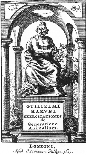
- ↑ Author includes this and the succeeding frontispiece from Harvey because they are supposed to come from a first edition. It is usually stated. however, that only one first edition appeared in London and three in Amsterdam during the same year. The phrase ex ovo omnia is found on the egg-shell in both. It is unknown who introduced these illustrations, but it is not improbable that it was the printer.
| Historic Disclaimer - information about historic embryology pages |
|---|
| Pages where the terms "Historic" (textbooks, papers, people, recommendations) appear on this site, and sections within pages where this disclaimer appears, indicate that the content and scientific understanding are specific to the time of publication. This means that while some scientific descriptions are still accurate, the terminology and interpretation of the developmental mechanisms reflect the understanding at the time of original publication and those of the preceding periods, these terms, interpretations and recommendations may not reflect our current scientific understanding. (More? Embryology History | Historic Embryology Papers) |
Meyer AW. 1932 - Essays on the History of Embryology: Part I | Part II | Part III | Part IV | Part V | Part VI | Part VII | Part VIII | Part IX | Part X | Part XI | Arthur Meyer | Historic Embryology Papers
Cite this page: Hill, M.A. (2024, April 28) Embryology Meyer - Essays on the History of Embryology 4. Retrieved from https://embryology.med.unsw.edu.au/embryology/index.php/Meyer_-_Essays_on_the_History_of_Embryology_4
- © Dr Mark Hill 2024, UNSW Embryology ISBN: 978 0 7334 2609 4 - UNSW CRICOS Provider Code No. 00098G


