File:Xenopus cornea development timeline.jpg

Original file (1,288 × 800 pixels, file size: 77 KB, MIME type: image/jpeg)
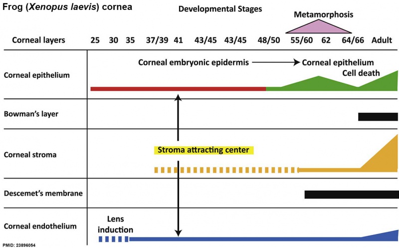
Xenopus cornea development timeline
Diagram summarizing the developmental timescale of all corneal layers. Thickness of the bars corresponds to the size of the tissue. Broken bar suggests periods of mesenchymal invasion in the formation of corneal stroma and endothelium.
- Links: Cornea Development | Frog Development
Reference
<pubmed>23896054</pubmed>| Exp Eye Res.
Copyright
Creative Commons Attribution License (CC BY)
This article is available under the terms of the Creative Commons Attribution License (CC BY). You may distribute and copy the article, create extracts, abstracts, and other revised versions, adaptations or derivative works of or from an article (such as a translation), to include in a collective work (such as an anthology), to text or data mine the article, including for commercial purposes without permission from Elsevier. The original work must always be appropriately credited.
Permission is not required for this type of reuse.
File history
Click on a date/time to view the file as it appeared at that time.
| Date/Time | Thumbnail | Dimensions | User | Comment | |
|---|---|---|---|---|---|
| current | 12:17, 6 September 2014 | | 1,288 × 800 (77 KB) | Z8600021 (talk | contribs) | ==Xenopus cornea development timeline== ===Reference=== <pubmed>23896054</pubmed>| [http://www.sciencedirect.com/science/article/pii/S0014483513002194 Exp Eye Res.] ====Copyright==== Creative Commons Attribution License (CC BY) This article is ava... |
You cannot overwrite this file.
File usage
The following 2 pages use this file: