File:Wislocki1920 plate 3.jpg

Original file (975 × 1,200 pixels, file size: 219 KB, MIME type: image/jpeg)
Plate 3
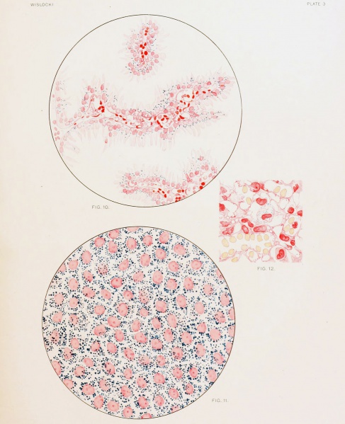
Fig. 10. Section of the endodermal villi, the modified remains of the "inverted yolk-sac," heavily laden with pigment after repeated injection of a colloidal dye into the maternal circulation.
Fig. 11. Wall of the "inverted yolk-sac," showing the endodermal cells as they appear 24 hours after an injection of trypan-blue into the amniotic cavity. Fresh membrane slightly eounterstained with alum carmine.
Fig. 12. Section through placenta of guinea-pig fetus measuring 40 mm., showing a vitally stained macrophage 26 hours after injection of trypan-blue into the amniotic cavity'.
| Historic Disclaimer - information about historic embryology pages |
|---|
 |
File history
Click on a date/time to view the file as it appeared at that time.
| Date/Time | Thumbnail | Dimensions | User | Comment | |
|---|---|---|---|---|---|
| current | 17:45, 27 December 2012 | | 975 × 1,200 (219 KB) | Z8600021 (talk | contribs) |
You cannot overwrite this file.
File usage
The following page uses this file: