File:Wheeler-plate02.jpg

Original file (829 × 1,115 pixels, file size: 197 KB, MIME type: image/jpeg)
Plate 2
Original plate images magnification shown in brackets.
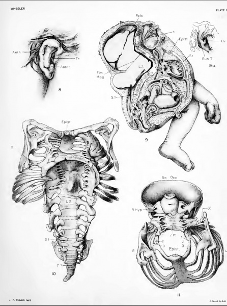
Fig. 8. Sketch of right ear
(natural size), showing the anthelix unusually prominent. The tragus lies relatively higher than normal, over rather than horizontally opposite the antitragus. The whole ear very narrow.
Fig. 9. Sagittal section
Main outlines were geometrically projected and detail drawn free-hand. The viscera retain approximately their normal position, absence of the soft palate is shown. The tip of the tongue lies over the left anlage of the spht uvula. The vertebral column is bent and shortened and irregularly fused in its upper part. The arches of all the vertebrse are lacking. A fibrous band lies over the upper sacral vertebra, joining the opposing defective arches in that region and forming a short spinal canal. The section passes to the left of the sella turcica. The falx cerebri is seen well over on left side. The outhne of the central nervous system, as is here shown, is used reversed for a diagram in figure 24. The section passes near the median margin of the left sac. (half original size)
Fig. 9a. Gives left side of bilateral anlage of uvula and orifice of eustachian tube
(Natural size)
Fig. 10. Shows a dorsal view of the mounted skeleton, with scapulae in place
Varying degrees of gaping vertebral arches are shown at different levels of the spinal column. In the cervical and thoracic regions defective vertebral arches are fused together and markedly everted. In the upper lumbar region they are individually distinct, but still widely everted, while in the lower lumbar and sacral regions they are distinct and bent toward one another. The lumbar transverse processes and the sacral lateral processes are well developed and the coccyx of four segments is seen bent well to the left. In the lower thoracic region a cartilaginous spur projects dorsa'vards from the vertebral bodies. All the thoracic and cervical vertebral bodies are fused together in a single plate. A sUght lateral bending in this plate is present. The foveal surfaces of the atlas face the reader. The intervertebral foramina show large spaces in the lumbar region, which are a sharp contrast to the tiny areas of the contracted thoracic intervertebral foramina. On the right, the rough surface of tho tip of the first lumbar arch is shown, which joins the occiput; and on I lie left, the second lumbar arch, whicli does the same. Crowding of the base of the ribs may be seen, including the first to the sixth on the right and the fifth to the ninth on the left. The sternum is considerably to the left of the milline. A persistent epistermmi is i)resent as a small cartilaginous knob, surrounding the manubrium. The irregular vertebral and superior margins of the scapula; are shown. On the left side the spicule of bone passes from the thoracic and cervical arches to the scapula. (Natural size).
Fig. 11. The superior view of the thoracic skeleton and the anterior surface of the cervical vertebral iilate and of the occiput
In the cervical part, no vertebral bodies are distinct, but irregular radicular, and transverse processes project laterally from the central plate. The abnormal spicule of bone on the left side may be seen passing from the fused transverse processes to the left scapula. This view shows how the foveal surfaces of the atlas are shifted to the right in relation to their underlying transverse processes. The right fovea almost overlies the tip of the right transverse process, while the left fovea leaves the left transverse process uncovered. The left transverse process is bent up and joins the pars lateralis, thus forming a rather large foramen. The anterior surface of the occiput shows an asymmetrical oval outline pierced by a foramen, in its center. The double exit of the right hypoglosssal canal shows. The irregular superior margins of the scapula are seen. The episternum and the aborted second rib are demonstrated. (Natural size).
File history
Click on a date/time to view the file as it appeared at that time.
| Date/Time | Thumbnail | Dimensions | User | Comment | |
|---|---|---|---|---|---|
| current | 08:35, 16 February 2011 |  | 829 × 1,115 (197 KB) | S8600021 (talk | contribs) | ==Plate 2== Original plate images magnification shown in brackets. ===Fig. 8. Sketch of right ear=== (natural size), showing the anthelix unusually prominent. The tragus lies relatively higher than normal, over rather than horizontally opposite the antit |
You cannot overwrite this file.
File usage
The following page uses this file: