File:Waterston1917 plate02.jpg

Original file (1,280 × 1,485 pixels, file size: 257 KB, MIME type: image/jpeg)
Plate 2
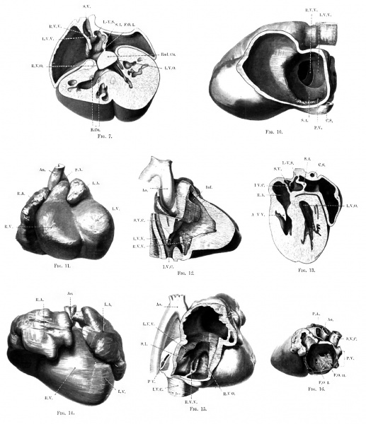
fig. 7. Caudal half of the same model divided horizontally.
fig. 10. View from the left of the same model after removal of the lateral wall of the left atrium.
fig. 11. Ventral aspect of model of 16-mm embryo.
fig. 12. The same model from the right side after removal of the lateral wall of the right atrium and ventricle, and of the right venous valve.
fig. 13. Caudal portion of model of heart of 20-mm embryo, divided horizontally.
fig. 14. Ventral aspect of model of 30-mm embryo.
fig. 15. Same model viewed from the right after removal of the lateral wall of the atrium and of the right venous valve.
fig. 16. Interior of the left atrium of heart of a fully developed child, with foramen ovale I persisting in addition to foramen ovale II.
| Figure Abbreviations |
|---|
|
A. = Bulbar cushion A. Ao. = Aorta. A.S. = Aortic sinus. Atr. = Atrium. A.-V.V. = Atrio-ventricular valve. A.-V.B. = Atrio-ventricular bundle. A.F. = Annulus fibrosus B.C. = Bulbus cordis. B.Cu. = Bulbar cushion B. = Bulbar cushion B. B.V. = Bulbo-ventricular. C.S. = Coronary sinus. E.T. = Endothelial tube. End. Cu. = Endocardial cushions of the atrial canal. F.O.1 and F.O. II = Foramen ovale primum and secundum Inf. = Infundibulum of right ventricle. I.V.C. = Inferior vena cava. I.-V.S. = Intersepto-valvular space. I.V.F. = Interventricular foramen. L.A. = Left atrium. R.A. = Right atrium. L.C. = Lateral cusp of atrio-ventricular valve. M.C. = Medial cusp of atrio-ventricular valve. L.V. = Left ventricle. L.A.-V.O. and R. A.-V.0. = Left and right atrio-ventricular openings. L.V.O. = Left venous ostium. R.V.O. = Right venous ostium. L.V.V. and R.V.V. = Left and right venous valves. O. = Opening of sinu-atrial chamber into the right atrium. P.A. = Pulmonary artery. P.V. = Pulmonary vein. RV. = Right ventricle. R.P.V. = Right pulmonary vein. S. I. = Septum primnm atriorum. S.-A.O. = Sinu-atrial orifice. S.B. = Septum of the bulbus cordis. S.V.= Sinus venosus. S.V.C. = Superior vena cava. Sin. At. = Sinu-atrial chamber. T.A. = Truncus arteriosus. T.V. = Tensor valvulae. V.Ao. = Ventral aorta. V.C.S. = Vena cava superior. |
Reference
Waterston D. The development of the heart in man. (1917) Trans. Roy. Soc. Edin., 7(2): 258-302.
Cite this page: Hill, M.A. (2024, April 26) Embryology Waterston1917 plate02.jpg. Retrieved from https://embryology.med.unsw.edu.au/embryology/index.php/File:Waterston1917_plate02.jpg
- © Dr Mark Hill 2024, UNSW Embryology ISBN: 978 0 7334 2609 4 - UNSW CRICOS Provider Code No. 00098G
File history
Click on a date/time to view the file as it appeared at that time.
| Date/Time | Thumbnail | Dimensions | User | Comment | |
|---|---|---|---|---|---|
| current | 10:32, 10 March 2017 |  | 1,280 × 1,485 (257 KB) | Z8600021 (talk | contribs) | |
| 10:30, 10 March 2017 |  | 2,408 × 3,010 (587 KB) | Z8600021 (talk | contribs) |
You cannot overwrite this file.
File usage
The following page uses this file: