File:Waterston1915 fig02.jpg
From Embryology
Size of this preview: 800 × 580 pixels. Other resolution: 923 × 669 pixels.
Original file (923 × 669 pixels, file size: 117 KB, MIME type: image/jpeg)
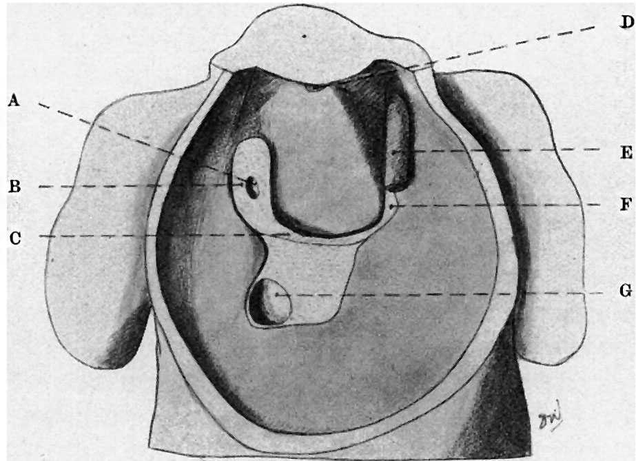
Fig. 2. Dorsal wall and floor of pericardial cavity of 6 mm Embryo
A, right duct of Cuvier; B, mesocardlum ; C, dorsal margin of septum trausversum; D, upper remains of dorsal mesocardium; E, left duct. of Cuvier ; F, mesocardial fold.
| Historic Disclaimer - information about historic embryology pages |
|---|
 |
Reference
Waterston D. Developmental changes in the pericardium, the mesocardia, and the pleural sacs in the human embryo. (1915) J Anat Physiol., 50(1): 24-9. PMID 17233049
Cite this page: Hill, M.A. (2024, April 26) Embryology Waterston1915 fig02.jpg. Retrieved from https://embryology.med.unsw.edu.au/embryology/index.php/File:Waterston1915_fig02.jpg
- © Dr Mark Hill 2024, UNSW Embryology ISBN: 978 0 7334 2609 4 - UNSW CRICOS Provider Code No. 00098G
File history
Click on a date/time to view the file as it appeared at that time.
| Date/Time | Thumbnail | Dimensions | User | Comment | |
|---|---|---|---|---|---|
| current | 17:05, 24 August 2015 | | 923 × 669 (117 KB) | Z8600021 (talk | contribs) | |
| 17:01, 24 August 2015 |  | 1,208 × 846 (222 KB) | Z8600021 (talk | contribs) |
You cannot overwrite this file.
File usage
The following page uses this file: