File:Uterine vascular anastomoses.jpg

Original file (1,200 × 370 pixels, file size: 48 KB, MIME type: image/jpeg)
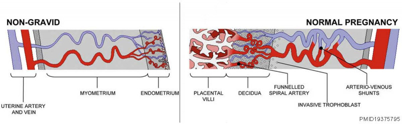
Uterine Vascular Anastomoses
Normal pregnancy is characterized by the formation of large anastomoses, arteriovenous shunts, that persist in the immediate post-partum period.
- Links: Figure - Uterine and placental vasculature | Uterine vascular anastomoses | Figure - Placenta spiral artery conversion | Placenta Development
Reference
Burton GJ, Woods AW, Jauniaux E & Kingdom JC. (2009). Rheological and physiological consequences of conversion of the maternal spiral arteries for uteroplacental blood flow during human pregnancy. Placenta , 30, 473-82. PMID: 19375795 DOI.
Copyright
© 2009 Elsevier Ltd. “This is an unofficial translation of an article that appeared in an Elsevier publication. Elsevier has not endorsed this translation.”
http://www.elsevier.com/wps/find/authorsview.authors/supplementalterms1.0
Original File name: Gr2.jpg cartoons cropped and relabelled from original figure.
Cite this page: Hill, M.A. (2024, April 25) Embryology Uterine vascular anastomoses.jpg. Retrieved from https://embryology.med.unsw.edu.au/embryology/index.php/File:Uterine_vascular_anastomoses.jpg
- © Dr Mark Hill 2024, UNSW Embryology ISBN: 978 0 7334 2609 4 - UNSW CRICOS Provider Code No. 00098G
File history
Click on a date/time to view the file as it appeared at that time.
| Date/Time | Thumbnail | Dimensions | User | Comment | |
|---|---|---|---|---|---|
| current | 12:24, 4 July 2018 | 1,200 × 370 (48 KB) | Z8600021 (talk | contribs) |
You cannot overwrite this file.
File usage
The following page uses this file: