File:Stricht-plate03.jpg
Original file (1,023 × 1,260 pixels, file size: 230 KB, MIME type: image/jpeg)
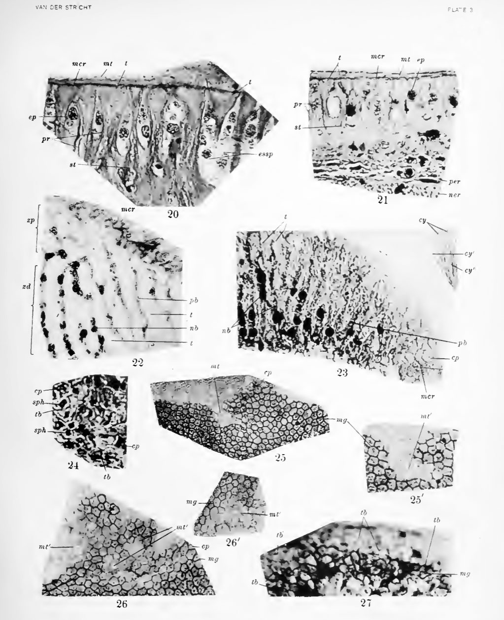
Plate 3
20. Photograph of section vertical to surface of crista spiralis. Pig embryo 190 mm. Bouin's fluid; Mallory's stain.
21. Photograph of section vertical to surface of crista spiralis. Young dog, age 4 months. Trichloracetic acid: iron hematoxylin, Stain - Congo red, light green.
22. Photograph of section tangential to surface of crista spiralis. Pig embryo 190 mm. Trichloracetic acid; iron hematoxylin, Stain - Congo red.
23. Photograph of section tangential to surface of crista spiralis in adult bat (Vesperlilw fuscus). Zenker's fluid, iron hematoxylin, Stain - Congo red
24. Photograph of section tangential to surface of crista spiralis. Young dog. Bouin's fluid; iron hematoxylin; Stain - Congo red.
25, 25', 26, 26'. Photographs of sections tangential to surface of greater ridge. New-born dog. Trichloracetic acid; iron hematoxylin, Stain - Congo red.
27. Photograph of section tangential to surface of greater ridge. Pig embryo 93.5 mm. Fixation by uranium nitrate method of Ramon y Cajal.
- Stricht Links: Plate 1 | Plate 2 | Plate 3 | Plate 4 | Membrana Tectoria and the Crista Spiralis | Contributions to Embryology Series
| Historic Disclaimer - information about historic embryology pages |
|---|
 |
Reference
O. Van der Stricht. The Genesis and Structure of the Membrana Tectoria and the Crista Spiralis of the Cochlea. Contributions to Embryology (1918) Vol. 7 pp55- 86.
Cite this page: Hill, M.A. (2024, April 26) Embryology Stricht-plate03.jpg. Retrieved from https://embryology.med.unsw.edu.au/embryology/index.php/File:Stricht-plate03.jpg
- © Dr Mark Hill 2024, UNSW Embryology ISBN: 978 0 7334 2609 4 - UNSW CRICOS Provider Code No. 00098G
File history
Click on a date/time to view the file as it appeared at that time.
| Date/Time | Thumbnail | Dimensions | User | Comment | |
|---|---|---|---|---|---|
| current | 08:56, 7 April 2011 | | 1,023 × 1,260 (230 KB) | S8600021 (talk | contribs) | ==Plate 3== 20. Photograph of section vertical to surface of crista spiralis. Pig embryo 190 mm. Bouin's fluid; Mallory's stain. 21. Photograph of section vertical to surface of crista spiralis. Young dog, age 4 months. Trichloracetic acid: iron hemato |
You cannot overwrite this file.
File usage
The following page uses this file: