File:Streeter1917 fig02.jpg

Original file (1,000 × 629 pixels, file size: 158 KB, MIME type: image/jpeg)
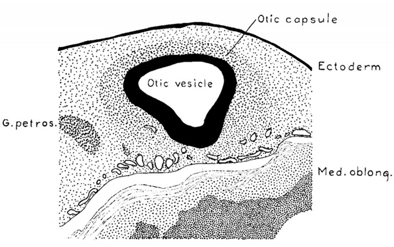
Fig. 2. Section through the region of the otic vesicle in a human embryo 9 mm long
(Carnegie Collection No. 721) enlarged 66.6 diameters. The primordial of the otic capsule, consisting of condensed mesenchyme, can be seen enclosing the vesicle on its lateral surface.
A definite layer of such nuclei is not found until the embryo reaches a length of about 9 mm.; it is then possible to recognize a fairly Well outlined zone of mesenchyme which represents the otic capsule in its first stage of development. In figure 2 is shown a sketch indicating the relations which exist at that time. It represents a transverse section through the otic vesicle at the level of the attachment of the endolymphatic appendage. The zone of condensed mesenchyme forming the primordium of the otic capsule abuts directly against the lateral wall of the vesicle and extends from there to a point about one-half the distance between the vesicle and the ectoderm. On the median side of the vesicle this zone is lacking, although there is a considerable number of mesenchyme.cel1s clustered around the vascular plexus ensheathing the central nervous system, and among the nerve rootlets of the acoustic complex. When this zone is analyzed under higher magnification it is found that it still consists essentially of a mesenchymal syncytium.
| Historic Disclaimer - information about historic embryology pages |
|---|
| Pages where the terms "Historic" (textbooks, papers, people, recommendations) appear on this site, and sections within pages where this disclaimer appears, indicate that the content and scientific understanding are specific to the time of publication. This means that while some scientific descriptions are still accurate, the terminology and interpretation of the developmental mechanisms reflect the understanding at the time of original publication and those of the preceding periods, these terms, interpretations and recommendations may not reflect our current scientific understanding. (More? Embryology History | Historic Embryology Papers) |
- Links: Fig 1 | Fig 2 | Fig 3 | Fig 4 | Fig 5 | Fig 6 | Fig 7 | Fig 8 | Fig 9 | Fig 10 | Streeter 1917 | Historic Embryology Papers | Carnegie Embryos
Reference
Streeter GL. The factors involved in the excavation of the cavities in the cartilaginous capsule of the ear in the human embryo. (1917) Amer. J Anat. 22: 1–25.
Cite this page: Hill, M.A. (2024, April 26) Embryology Streeter1917 fig02.jpg. Retrieved from https://embryology.med.unsw.edu.au/embryology/index.php/File:Streeter1917_fig02.jpg
- © Dr Mark Hill 2024, UNSW Embryology ISBN: 978 0 7334 2609 4 - UNSW CRICOS Provider Code No. 00098G
File history
Click on a date/time to view the file as it appeared at that time.
| Date/Time | Thumbnail | Dimensions | User | Comment | |
|---|---|---|---|---|---|
| current | 08:31, 30 October 2015 |  | 1,000 × 629 (158 KB) | Z8600021 (talk | contribs) | |
| 08:30, 30 October 2015 |  | 1,300 × 992 (317 KB) | Z8600021 (talk | contribs) | {{Streeter1917a}} |
You cannot overwrite this file.
File usage
The following page uses this file:
