File:Streeter002-3.jpg
Original file (607 × 800 pixels, file size: 106 KB, MIME type: image/jpeg)
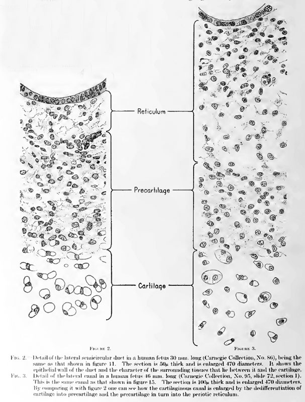
Fig. 2. Detail of the lateral semicircular duct in a human fetus 30 mm. long
(Carnegie Collection, No. 86), being the same as that shown in figure 11. The section is 50 micron thick and is enlarged 470 diameters.
It shows the epithelial wall of the duct and the character of the surrounding tissues that lie between it and the cartilage.
Fig. 3. Detail of the lateral canal in a human fetus 16 mm. long
(Carnegie Collection, No. 95, slide 72, section 1). This is the same canal as that shown in figure 15. The section is 100 micron thick and is enlarged 470 diameters.
By comparing it with figure 2 one can see how the cartilaginous canal is enlarged by the dedifferentiation of cartilage into precartilage and the precartilage in turn into the periotic reticulum.
Reference
Streeter G.L. The histogenesis and growth of the otic capsule and its contained periotic tissue-spaces in the human embryo Contributions to Embryology Carnegie Institution No.20 (1918) pp5-54, 4 text-figures and 4 plates.
File history
Click on a date/time to view the file as it appeared at that time.
| Date/Time | Thumbnail | Dimensions | User | Comment | |
|---|---|---|---|---|---|
| current | 00:13, 15 February 2011 | | 607 × 800 (106 KB) | S8600021 (talk | contribs) |
You cannot overwrite this file.
File usage
The following 2 pages use this file: