File:Stewart1955 plate02.jpg
Original file (1,280 × 1,036 pixels, file size: 134 KB, MIME type: image/jpeg)
Plate 2
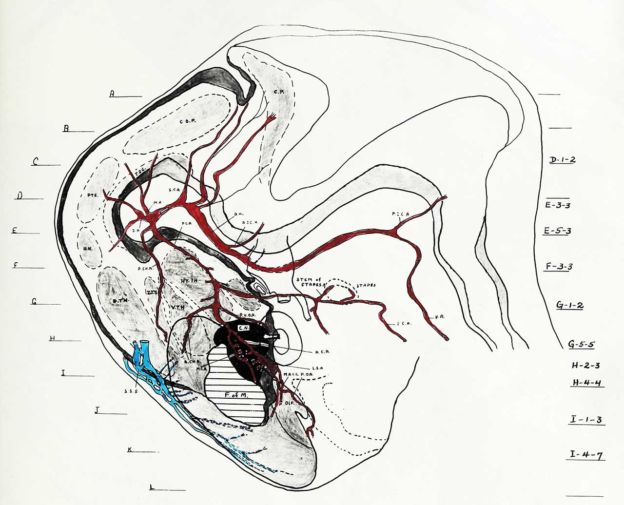
Plate II is a graphic reconstruction of the nuclear configurations forming the medial wall of the mesencephalon and diencephalon of the right half of a 12.5 mm., C-R length, human Embryo's brain, with the left cerebral hemisphere and the arterial and venous systems of the left side superimposed. Reconstruction of this type was used in order to illustrate, in two dimensions, the three dimensional aspect of the erabxyo brain. Serial cross sections were used to reconstruct structures lying lateral to the medial wall, with special reference to the blood vessels. Some of these sections are photographically reproduced in Figures A1 to A10 and are labelled on plate II, on the left, as planes A-L. The numbering of these planes, on the right, refers to the original sections.
Plates III and IV are enlarged reproductions of the areas outlined on the onion paper on plate II.
NUCLEI:
Plate II: Both diencephalon and telencephalon show the same nuclear structure as depicted in plate I but in a less expanded state.
| Abbreviations | |
|---|---|
| A.C.A. anterior cerebral artery
A.CH.A. anterior choroidal artery A.CO.A. anterior communicating artery A. COM. anterior commissure A.P. anterior plexus B.A. basilar artery CER.E. cerebral hemisphere C.N. corpus striatum (caudate nucleus) CO.P. collicular plate C.P. cerebellar plate D.A. diencephalic artery D.C.A. diencephalic artery DIM. diencephalon D.TH. dorsal thalamus F. of M. foramen of Monro HIPPO.P. hippocampus primordium H.N. habenular nucleus HYP.A.L. hypophysis anterior lobe HYPOPH. hypophysis HY.TH. hypothalamus I.G. internal capsule I.C.A. Internal carotid artery I.C.V. internal cerebral vein. IT.S. intrathalamic sulcus L.G.B. lateral geniculate boc^r L.S.A. lateral striate arteries M.A. mesencephalic artery M.A.C.C. medial artery of the corpus sallosum M.C.A. middle cerebral artery MES. mesencephalon M.R. mammalary recess M.S.A. medial striate artery (Heubner's recurrent) |
O.A. ophthalmic artery
O.G. optic groove OLF.N. olfactory nerve OLF.TUB. olfactory tubercle Q.N. optic nerve O.R. optic recess O x putamen PA. pallium P.C. posterior commissure PC. A. posterior communicating artery P.CO.A. posterior communicating artery P.CH.A. posterior choroidal artery P.I.C.A. posterior inferior cerebellar artery PIT. pituitary gland primordium P.O.A. primitive olfactory artery PO.R. pre-optic recess PTE. pretecturn RHOMB. rhombencephalon 5.C.A. superior cerebellar artery S.D.TH. dorsal thalamic sulcus S.M.TH. medial thalamic sulcus S.O.N. supra-optic nucleus S.P.C.A. superior portion of the posterior cerebral artery s.s. straight sinus S.S.P. superior sagittal sinus STAPES.A. stapedial artery S.V.TH. ventral thalamic sulcus TEG. tegmentum V.A. vertebral artery VEL. INTER. velum interpositum V.S. ventricular sulcus V.TH. ventral thalamus Z.IT. zona intrathamica |
Reference
Stewart GG. The development of the blood supply to the human embryo basal ganglia. (1955) University of Alberta, Canada.
Cite this page: Hill, M.A. (2024, April 25) Embryology Stewart1955 plate02.jpg. Retrieved from https://embryology.med.unsw.edu.au/embryology/index.php/File:Stewart1955_plate02.jpg
- © Dr Mark Hill 2024, UNSW Embryology ISBN: 978 0 7334 2609 4 - UNSW CRICOS Provider Code No. 00098G
File history
Click on a date/time to view the file as it appeared at that time.
| Date/Time | Thumbnail | Dimensions | User | Comment | |
|---|---|---|---|---|---|
| current | 09:49, 11 June 2018 | | 1,280 × 1,036 (134 KB) | Z8600021 (talk | contribs) | |
| 09:48, 11 June 2018 |  | 3,047 × 3,648 (747 KB) | Z8600021 (talk | contribs) |
You cannot overwrite this file.
File usage
The following page uses this file: