File:Stage7-sem5.jpg
Original file (1,000 × 735 pixels, file size: 202 KB, MIME type: image/jpeg)
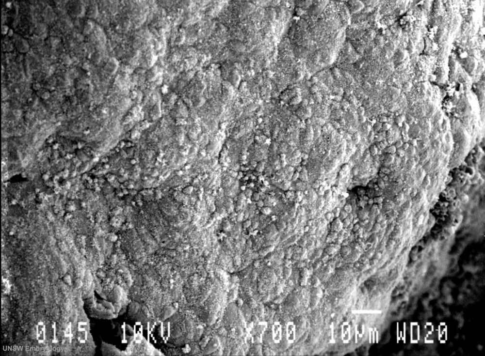
Human Embryo Carnegie stage 7 - Primitive Streak
17 days, pre-somite, scanning electron micrograph image
Selected region of embryonic disc (epiblast/ectoderm layer) dorsal view, with amniotic membrane partially removed.
Primitive node (Henson's node) in centre of disc and primitive streak, shown as indentation in disc is extending to the left.
Scale bar 10 microns
See also: Stage7-sem4.jpg image cropped and new scale bar added | Stage7-sem3.jpg original image
Image Source: Scanning electron micrographs of the Carnegie stages of the early human embryos are reproduced with the permission of Prof Kathy Sulik, from embryos collected by Dr. Vekemans and Tania Attié-Bitach. Images are for educational purposes only and cannot be reproduced electronically or in writing without permission.
Cite this page: Hill, M.A. (2024, April 26) Embryology Stage7-sem5.jpg. Retrieved from https://embryology.med.unsw.edu.au/embryology/index.php/File:Stage7-sem5.jpg
- © Dr Mark Hill 2024, UNSW Embryology ISBN: 978 0 7334 2609 4 - UNSW CRICOS Provider Code No. 00098G
File history
Click on a date/time to view the file as it appeared at that time.
| Date/Time | Thumbnail | Dimensions | User | Comment | |
|---|---|---|---|---|---|
| current | 13:36, 21 August 2009 | | 1,000 × 735 (202 KB) | MarkHill (talk | contribs) | Human Embryo Carnegie stage 7, 17 days, pre-somite, scanning electron micrograph image Selected region of embryonic disc (epiblast/ectoderm layer) dorsal view, with amniotic membrane partially removed. Primitive node (Henson's node) in centre of disc a |
You cannot overwrite this file.
File usage
The following 3 pages use this file: