File:Spleen histology 07.jpg
From Embryology
Size of this preview: 750 × 600 pixels. Other resolution: 1,000 × 800 pixels.
Original file (1,000 × 800 pixels, file size: 251 KB, MIME type: image/jpeg)
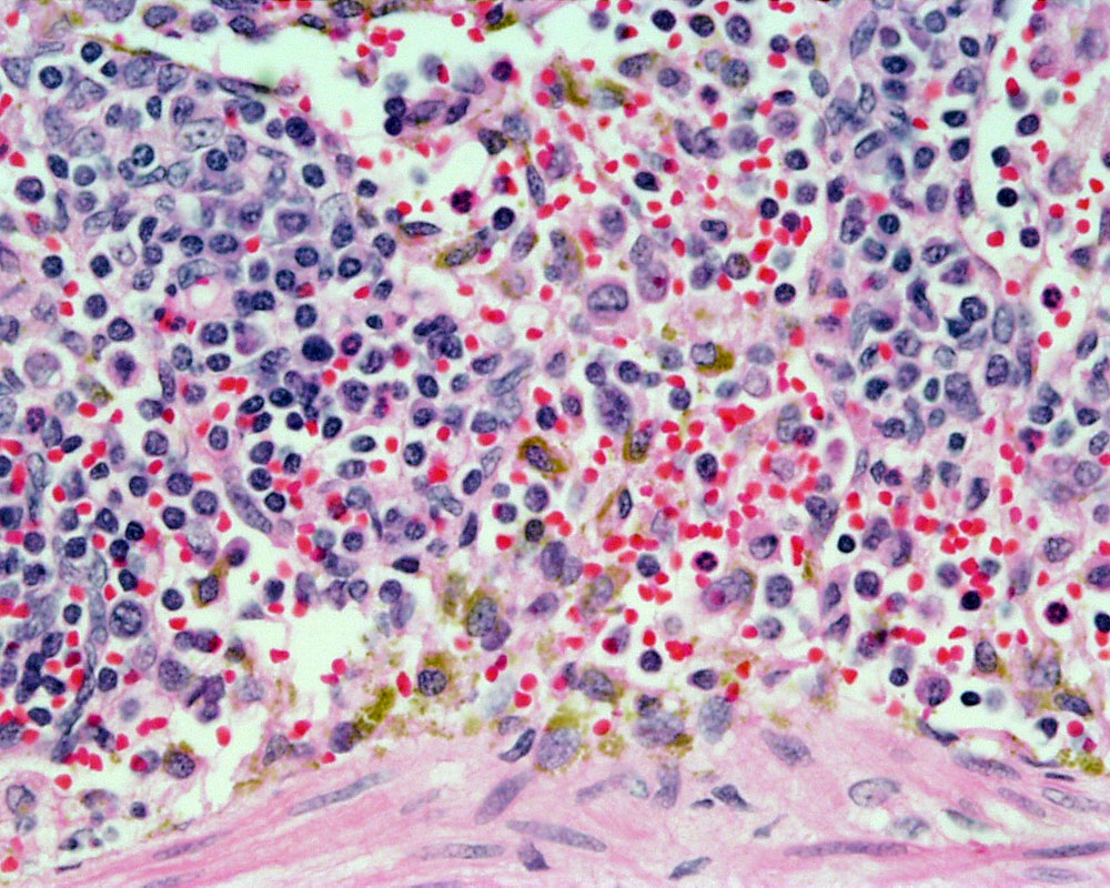
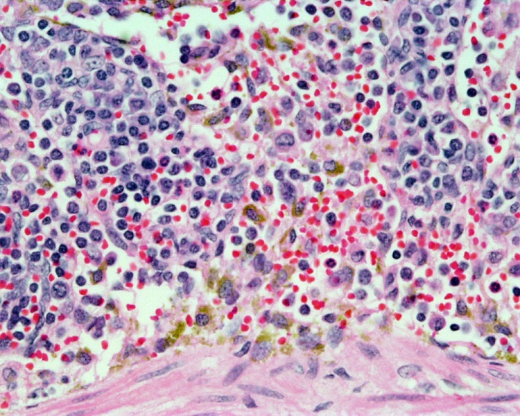
Spleen Histology
(Stain - Haematoxylin Eosin)
- Human spleen, lymphoid tissue, red pulp, splenic sinuses, splenic cords,
- macrophages shown in yellow/brown.
Links: Histology | Histology Stains | Blue Histology images copyright Lutz Slomianka 1998-2009. The literary and artistic works on the original Blue Histology website may be reproduced, adapted, published and distributed for non-commercial purposes. See also the page Histology Stains.
Cite this page: Hill, M.A. (2024, April 26) Embryology Spleen histology 07.jpg. Retrieved from https://embryology.med.unsw.edu.au/embryology/index.php/File:Spleen_histology_07.jpg
- © Dr Mark Hill 2024, UNSW Embryology ISBN: 978 0 7334 2609 4 - UNSW CRICOS Provider Code No. 00098G
Original file name: Spl40he.jpg
File history
Click on a date/time to view the file as it appeared at that time.
| Date/Time | Thumbnail | Dimensions | User | Comment | |
|---|---|---|---|---|---|
| current | 17:44, 21 February 2011 | | 1,000 × 800 (251 KB) | S8600021 (talk | contribs) | ==Human Spleen== Lymphoid tissue, red pulp, splenic sinuses, splenic cords, macrophages. Stain: H&E Original file name: Spl40he.jpg {{Blue Histology}} Category:Human Category:Spleen Category:Endocrine Category:Histology [[Category:Im |
You cannot overwrite this file.