File:Spaulding-fig12.jpg
Original file (445 × 607 pixels, file size: 25 KB, MIME type: image/jpeg)
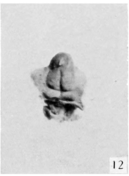
Fig. 12. Carnegie Embryo No. 1750
17.2 mm., female. X 6
Stage 5, 16 to 17 mm. (fig. 8, male; fig. 12, female). By the time the embryo has attained a length of 16 to 17 mm. the genital tubercle has elongated into a narrow, conical organ which, because of its modified shape and its separation from . the surrounding body areas by the newly formed labio-scrotal swellings, will now be called the phallus. The male embryo representing this stage (fig. 8) shows the phallus as more nearly cylindrical than in any of the younger embryos, the lateral buttresses having, to a marked extent, merged into its body. The groove limiting the glans is present, although not clearly shown in the photograph. The urogenital opening is a narrow orifice extending almost the full length of the phallus, limited distally by a pronounced epithelial tag. The urethral folds are considerably broader than in any of the younger stages, their outer margins being sharply separated from the remains of the lateral buttresses by a concave depression. The most pronounced change, however, is the presence of the labio-scrotal swellings as a pair of distinct, rounded ridges, one on each side of the base of the phallus and separated from it by a broad lateral phallic groove. As regards this feature, it is quite likely that this embryo represents a slightly older stage, as the male specimen of the next stage shows more clearly the probable beginnings of these swellings, and the female selected as the counterpart for this stage does not show them to any marked degree, if they are present at all. It is, of course, very possible that there is normally more or less variation in the time of development of these structures, and that this embryo (fig. 8) is merely slightly precocious in this respect.
| Historic Disclaimer - information about historic embryology pages |
|---|
 |
- Figure Links: Text | Text Figure 1 | Text Figure 2 | Plate 1 | Fig. 1 | Fig. 2 | Fig. 3 | Fig. 4 | Fig. 5 | Fig. 6 | Plate 2 | Fig. 7 | Fig. 8 | Fig. 9 | Fig. 10 | Fig. 11 | Fig. 12 | Fig. 13 | Fig. 14 | Fig. 15 | Fig. 16 | Fig. 17 | Fig. 18 | Fig. 19 | Fig. 20 | Fig. 21 | Fig. 22 | Plate 3 | Fig. 23 | Fig. 24 | Fig. 25 | Fig. 26 | Fig. 27 | Fig. 28 | Fig. 29 | Plate 4 | Fig. 30 | Fig. 31 | Fig. 32 |Fig. 33 | Fig. 34 | Fig. 35 | Fig. 36 | Fig. 37 | Fig. 38 | Fig. 39 | Fig. 40 | Fig. 41 | Fig. 42 | Fig. 43 | Fig. 44 | Fig. 45 | Fig. 46 | Fig. 47 | Fig. 48 | Fig. 49 | Fig. 50 | Fig. 51 | Fig. 52 | Fig. 53 | Fig. 54
| Historic Disclaimer - information about historic embryology pages |
|---|
|
Reference
Spaulding MH. The development of the external genitalia in the human embryo. (1921) Contrib. Embryol., Carnegie Inst. Wash. Publ. 81, 13: 69 – 88.
Cite this page: Hill, M.A. (2024, April 26) Embryology Spaulding-fig12.jpg. Retrieved from https://embryology.med.unsw.edu.au/embryology/index.php/File:Spaulding-fig12.jpg
- © Dr Mark Hill 2024, UNSW Embryology ISBN: 978 0 7334 2609 4 - UNSW CRICOS Provider Code No. 00098G
File history
Click on a date/time to view the file as it appeared at that time.
| Date/Time | Thumbnail | Dimensions | User | Comment | |
|---|---|---|---|---|---|
| current | 00:15, 15 April 2015 | | 445 × 607 (25 KB) | Z8600021 (talk | contribs) |
You cannot overwrite this file.
File usage
The following 3 pages use this file: