File:Smellie1754 table01.jpg

Original file (990 × 1,034 pixels, file size: 220 KB, MIME type: image/jpeg)
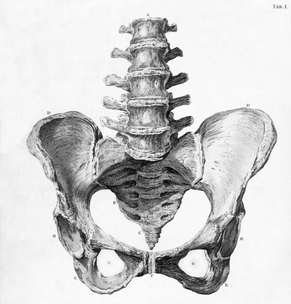
Table 1 The Bones of a well-formed Pelvis (Front view)
| In this Table, besides the general structure and figure of the several bones, the dimensions of the brim of the pelvis, and the distance between the under parts of the ossa ischium, are particularly to be attended to ; from which it will appear, that the cavity of the brim is commonly wider from side to side than from the back to the fore part, but that the sides below are in the contrary proportion. The reader, however, ought not to conclude, that every pelvis is similar in figure and dimensions, since even well-formed ones differ in some degree from each other. In general, the brim of the pelvis measures about five inches and a quarter from side to side, and four inches and a quarter from the back to the fore part ; there being likewise the same distance between the inferior parts of the ossa ischium. All these measures, however, must be understood as taken from the skeleton; for, in the subject, the cavity of the pelvis is considerably diminished by its teguments and contents. Correspondent also to this diminution, the usual dimensions of the head of the full-grown foetus are but three inches and a half from ear to ear, and four inches and a quarter from the fore to the hind head.
Vide Tab. XVI. XVII. XVIII. Also Vol. I. Chap. I. Sect. 1. 2. 3. where the form and dimensions of the pelvis, as well as of the head of the fetus, and the manner in which the same is protruded in labour through the basin, are fully treated of. Consult likewise Vol. II. Coll. 1. No. 1. 2. where cases are given of complaints of the pelvis arising from difficult labours. |
LegendA The five vertebra of the loins. B The os sacrum. C The os coccygis. D The ossa ilium. E The ossa ischium. F The ossa pubis. G The, foramina magna. H The acetabula. I The brim of the pelvis, or that circumference of its cavity, which is described at the sides of the inferior parts of the osssa ilium, and at the back and fore parts by the superior parts of the ossa pubis and sacrum. |
Table 1 Links: Table 1 colour | Table 1 bw
| Historic Disclaimer - information about historic embryology pages |
|---|
 |
File history
Click on a date/time to view the file as it appeared at that time.
| Date/Time | Thumbnail | Dimensions | User | Comment | |
|---|---|---|---|---|---|
| current | 14:41, 9 November 2012 | | 990 × 1,034 (220 KB) | Z8600021 (talk | contribs) |
You cannot overwrite this file.
File usage
The following 2 pages use this file: