File:Shaw1932 fig02.jpg
Original file (1,000 × 653 pixels, file size: 49 KB, MIME type: image/jpeg)
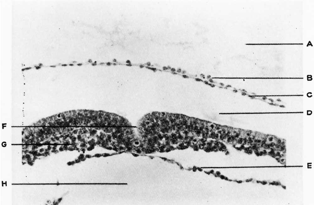
Fig. 2. Specimen No. 1. High power
Section through the region of primitive streak and groove. A, magma reticulare; B, extra-embryonic mesoderm; 0, cells of amnion; D, amniotic sac ; E, entoderm; F, primitive groove; G, embryonic mesoderm ; H, cavity of yolk sac.
Figs. 3 and 4. A, cerebral vesicle; B, heart; 0, coelom; D, intestine; E, primitive kidney; F, limb bud; G, dorsal aorta; H, neural canal; C, chorionic villi ; K, amnion; L, umbilical vessel ; M, mesone hric duct; N, neural canal of the embryo , 0, liver. The smaller embryo has no head, heart, or liver. The area of fusion is in the region of the primit ve kidney and umbilicus. The amnion is single.
Reference
Shaw W. Observations on two specimens of early human ova. (1932) Brit. Med.J., 1: 411-415.
Cite this page: Hill, M.A. (2024, May 9) Embryology Shaw1932 fig02.jpg. Retrieved from https://embryology.med.unsw.edu.au/embryology/index.php/File:Shaw1932_fig02.jpg
- © Dr Mark Hill 2024, UNSW Embryology ISBN: 978 0 7334 2609 4 - UNSW CRICOS Provider Code No. 00098G
File history
Click on a date/time to view the file as it appeared at that time.
| Date/Time | Thumbnail | Dimensions | User | Comment | |
|---|---|---|---|---|---|
| current | 17:06, 15 November 2017 | | 1,000 × 653 (49 KB) | Z8600021 (talk | contribs) | |
| 17:06, 15 November 2017 | 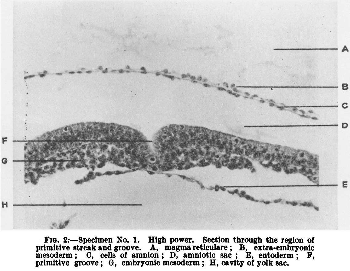 | 1,157 × 896 (103 KB) | Z8600021 (talk | contribs) |
You cannot overwrite this file.
File usage
The following page uses this file: