File:Sabin1915 plate05.jpg

Original file (2,861 × 2,231 pixels, file size: 1,013 KB, MIME type: image/jpeg)
Plate 5
Fig. 11. Dissection of an embryo pig 20 mm. long in which the vascular system has been injected with India ink through the umbilical vein. All of the viscera have been removed. X20.
A., aorta; v. A., v. azygos; v. B., common stem of the v. fibularis primitiva and the v. caudalis which becomes the lower segment of the vena cava; v. o. p., v. cardinalis posterior; v. L. T., v. lumbalis transversa; v. s. 2, v. spinalis of the second segment; v. TE., v. thoraco-epigastrica; v. u. P., v. ulnaris primitiva.
Fig. 12. Transverse section of an embryo pig 20 mm. long in which the vascular system has been injected with India ink through the umbilical artery. The section is to show the prevertebral plexus and passes through the median mesonephritic vein. The section is unstained and is 250 n thick. X77.
A., aorta; A. s., a. spinalis, which is now a branch of a single dorsal segmental artery; p. P., prevertebral plexus at the position of the future azygos vein, the limit of the development of the v. azygos for this stage being shown in fig. 11; p. p'., prevertebral plexus at the point in which the third segment of the inferior vena cava and the corresponding ascending lumbar vein develop at the level below the median mesonephritic vein; p. p. v., prevertebral plexus of small veins resting on the vertebra; v. M. M., v. mediana mesonephritica, the anastomosis across the mid-line of the embryo of the two vv. cardinalis mesiales; v. T. M., v. transversa mesialis of the Wolffian body.
Sabin 1915: plate 1 | plate 2 | plate 3 | plate 4 | plate 5 | plate 6 | plate 7 | pig
- Pig posterior cardinal veins
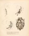
plate 1
plate 2
plate 3
plate 4
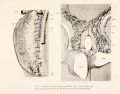
plate 5
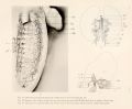
plate 6
plate 7


| Historic Disclaimer - information about historic embryology pages |
|---|
 |
References
Sabin FR. On the fate of the posterior cardinal veins and their relation to the development of the vena cava and azygos in the embryo pig. (1915) Pub. No. 223 Contrib. Embryol., Carnegie Inst. Wash. 3(7): 5-32. PDF
Cite this page: Hill, M.A. (2024, April 26) Embryology Sabin1915 plate05.jpg. Retrieved from https://embryology.med.unsw.edu.au/embryology/index.php/File:Sabin1915_plate05.jpg
- © Dr Mark Hill 2024, UNSW Embryology ISBN: 978 0 7334 2609 4 - UNSW CRICOS Provider Code No. 00098G
File history
Click on a date/time to view the file as it appeared at that time.
| Date/Time | Thumbnail | Dimensions | User | Comment | |
|---|---|---|---|---|---|
| current | 14:37, 30 July 2019 | | 2,861 × 2,231 (1,013 KB) | Z8600021 (talk | contribs) | from original scan |
| 12:24, 30 July 2019 |  | 752 × 581 (82 KB) | Z8600021 (talk | contribs) |
You cannot overwrite this file.
File usage
The following 11 pages use this file:
- Embryology History - Florence Sabin
- Paper - On the fate of the posterior cardinal veins and their relation to the development of the vena cava and azygos in the embryo pig (1915)
- File:Sabin1915 plate01.jpg
- File:Sabin1915 plate02.jpg
- File:Sabin1915 plate03.jpg
- File:Sabin1915 plate04.jpg
- File:Sabin1915 plate05.jpg
- File:Sabin1915 plate06.jpg
- File:Sabin1915 plate07.jpg
- Template:Ref-Sabin1915 figures
- Template:Sabin1915 plates gallery