File:Sabin1909 fig12.jpg
From Embryology

Size of this preview: 426 × 599 pixels. Other resolution: 713 × 1,003 pixels.
Original file (713 × 1,003 pixels, file size: 89 KB, MIME type: image/jpeg)
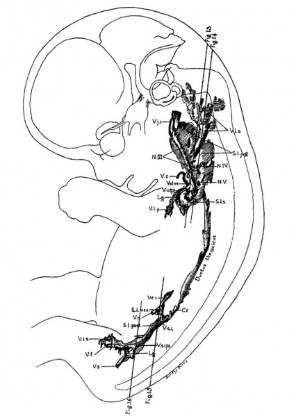
Fig 12. Human embryo measuring 30 mm
Fig. 12. Flat reconstruction of the primitive lymphatic system in a human embryo, 30 mm. long, Mall collection. No. 86. X about 5.4.
Legend
- C. c., clsterna chyli
- L. G, lymphoglandula : N. III., N. IV., and N. V.. Nn. cervlcales
- S. l. jug., saccus lymphatlcus jugularis
- S. l. mes., saculus lymphaticus mesentericus
- S. l. post., saccus lymphaticus posterior
- S. l. s.. ssccus lgmphatlcus subclavius
- V. c., vena cephallca ; $. c. l., vena cava inferior
- V. f., vena femoralls
- V. j. i. vena jugular18 inferior
- V. l. p., vasa lymphatica profunds
- V. l. s., vasa lymphatlca superficlalls
- V. r., vena renalis
- v. s., vena sciatica
- V. u. (p.), vena ulnarls (prlmltlva).
Approximately from above drawing and description.
- Links: Immune System Development
Reference
Sabin FR. The lymphatic system in human embryos, with a consideration of the morphology of the system as a whole. (1909) Amer. J Anat. 9(1): 43–91.
Cite this page: Hill, M.A. (2024, April 26) Embryology Sabin1909 fig12.jpg. Retrieved from https://embryology.med.unsw.edu.au/embryology/index.php/File:Sabin1909_fig12.jpg
- © Dr Mark Hill 2024, UNSW Embryology ISBN: 978 0 7334 2609 4 - UNSW CRICOS Provider Code No. 00098G
File history
Click on a date/time to view the file as it appeared at that time.
| Date/Time | Thumbnail | Dimensions | User | Comment | |
|---|---|---|---|---|---|
| current | 13:41, 30 March 2011 |  | 713 × 1,003 (89 KB) | S8600021 (talk | contribs) | ==Fig 12. Human embryo== Approximately Carnegie stage 21 from above drawing and description. ==Reference== Florence R. Sabin, The lymphatic system in human embryos, with a consideration of the morphology of the system as a whole |
You cannot overwrite this file.
File usage
The following page uses this file: