File:Placenta percreta 05.jpg

Original file (900 × 676 pixels, file size: 77 KB, MIME type: image/jpeg)
Placenta Percreta
Chorionic villi in the myometrium of uterus, which explain that the placenta percreta is noted at optical microscopy.
Reference
Ambrogi G, Ambrogi G & Marchi AA. (2018). Placenta Percreta and Uterine Rupture in the First Trimester of Pregnancy. Case Rep Obstet Gynecol , 2018, 6842892. PMID: 29850318 DOI.
Copyright
Copyright © 2018 Gabriel Ambrogi et al. This is an open access article distributed under the Creative Commons Attribution License, which permits unrestricted use, distribution, and reproduction in any medium, provided the original work is properly cited.
CRIOG2018-6842892.003.jpg
Cite this page: Hill, M.A. (2024, April 26) Embryology Placenta percreta 05.jpg. Retrieved from https://embryology.med.unsw.edu.au/embryology/index.php/File:Placenta_percreta_05.jpg
- © Dr Mark Hill 2024, UNSW Embryology ISBN: 978 0 7334 2609 4 - UNSW CRICOS Provider Code No. 00098G
File history
Click on a date/time to view the file as it appeared at that time.
| Date/Time | Thumbnail | Dimensions | User | Comment | |
|---|---|---|---|---|---|
| current | 14:46, 4 June 2018 | | 900 × 676 (77 KB) | Z8600021 (talk | contribs) | |
| 14:41, 4 June 2018 | 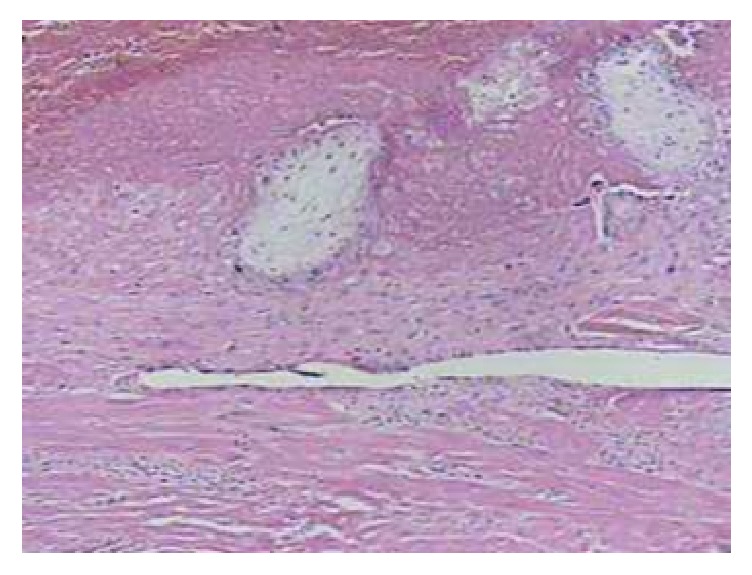 | 752 × 575 (110 KB) | Z8600021 (talk | contribs) | ==Placenta Percreta== Chorionic villi in the myometrium of uterus, which explain that the placenta percreta is noted at optical microscopy. CRIOG2018-6842892.003.jpg |
You cannot overwrite this file.
File usage
There are no pages that use this file.