File:Patten020.jpg

Original file (796 × 1,020 pixels, file size: 128 KB, MIME type: image/jpeg)
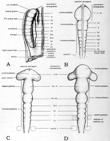
Fig. 20. Diagrams to show the neuromeric enlargements in the brain region of the neural tube
(Based on figures by Hill.)
A lateral view of neural plate from dissection of chick of 4 somites (24 hours)
B dorsal view of brain dissected out of 7-somite (26 to 27 hour) embryo
C dorsal view of brain from 10-somite (30 hour) embryo
D dorsal view of brain from 14-somite (36 hour) embryo
By about 33 hours of incubation the optic vesicles are established as paired lateral outgrowths of the prosencephalon. They soon extend to occupy the full width of the head (Fig. 20, C and Fig. 21). The distal portion of each of the vesicles thus comes to lie closely approximated to the superficial ectoderm, a relationship of importance in their later development. At first the cavities of the optic vesicles (opticoeles) are broadly confluent with the cavity of the prosencephalon (prosocoele). Somewhat later constrictions appear which mark more definitely the boundaries between the optic vesicles and the prosencephalon (Fig. 20, D and Fig. 22).
- Links: Introduction | Gametes and Fertilization | Segmentation | Entoderm | Primitive Streak and Mesoderm | Primitive Streak to Somites | 24 Hours | 24 to 33 Hours | 33 to 39 Hours | 40 to 50 Hours | Extra-embryonic Membranes | 50 to 55 Hours | Day 3 to 4 | References | All Figures
| Historic Disclaimer - information about historic embryology pages |
|---|
| Pages where the terms "Historic" (textbooks, papers, people, recommendations) appear on this site, and sections within pages where this disclaimer appears, indicate that the content and scientific understanding are specific to the time of publication. This means that while some scientific descriptions are still accurate, the terminology and interpretation of the developmental mechanisms reflect the understanding at the time of original publication and those of the preceding periods, these terms, interpretations and recommendations may not reflect our current scientific understanding. (More? Embryology History | Historic Embryology Papers) |
Reference
Patten BM. The Early Embryology of the Chick. (1920) Philadelphia: P. Blakiston's Son and Co.
Cite this page: Hill, M.A. (2024, April 26) Embryology Patten020.jpg. Retrieved from https://embryology.med.unsw.edu.au/embryology/index.php/File:Patten020.jpg
- © Dr Mark Hill 2024, UNSW Embryology ISBN: 978 0 7334 2609 4 - UNSW CRICOS Provider Code No. 00098G
File history
Click on a date/time to view the file as it appeared at that time.
| Date/Time | Thumbnail | Dimensions | User | Comment | |
|---|---|---|---|---|---|
| current | 01:07, 17 January 2011 |  | 796 × 1,020 (128 KB) | S8600021 (talk | contribs) | ==Fig. 20. Diagrams to show the neuromeric enlargements in the brain region of the neural tube== (Based on figures by Hill.) A , lateral view of neural plate from dissection of chick of 4 somites (24 hours) B, dorsal view of brain dissected out of 7-so |
You cannot overwrite this file.
File usage
The following 2 pages use this file:
