File:Parker1874 plate28.jpg
Original file (1,028 × 1,394 pixels, file size: 192 KB, MIME type: image/jpeg)
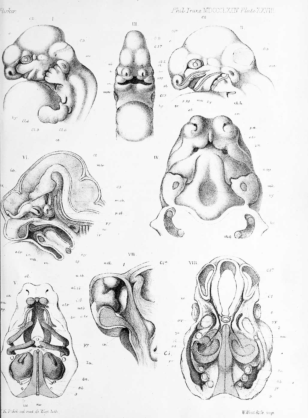
PLATE XXVIII.
First Stage. Emhryo Pig., f inch in length.
Fig. 1. Side view of upper part of embryo. X 7 diameters.
Fig. 2. A plan of the same, with facial arches. X 7 diameters.
Fig. 3. A front view of the same. X 7 diameters.
Fig. 4. A palatal view of the same, with the mandible and lower face removed. X 15 diameters.
Fig. 5. A plan of the skull and face, seen from below. X 10 diameters.
Fig. 6. A vertical section of the head. X 10 diameters.
Fig. 7. Part of the same, with median part of nasal region removed. X -0 diameters.
Fig. 8. Upper view of a horizontal section of the head, x 10 diameters.
Reference
Parker WK. On the structure and development of the skull in the pig (sus scrofa). (1874)
Cite this page: Hill, M.A. (2024, April 26) Embryology Parker1874 plate28.jpg. Retrieved from https://embryology.med.unsw.edu.au/embryology/index.php/File:Parker1874_plate28.jpg
- © Dr Mark Hill 2024, UNSW Embryology ISBN: 978 0 7334 2609 4 - UNSW CRICOS Provider Code No. 00098G
File history
Click on a date/time to view the file as it appeared at that time.
| Date/Time | Thumbnail | Dimensions | User | Comment | |
|---|---|---|---|---|---|
| current | 17:05, 8 May 2018 | | 1,028 × 1,394 (192 KB) | Z8600021 (talk | contribs) | |
| 17:03, 8 May 2018 |  | 2,808 × 3,808 (626 KB) | Z8600021 (talk | contribs) | PLATE XXVIII. First Stage. Emhryo Pig., f inch in length. Fig. 1. Side view of upper part of embryo. X 7 diameters. Fig. 2. A plan of the same, with facial arches. X 7 diameters. Fig. 3. A front view of the same. X 7 diameters. Fig. 4. A pala... |
You cannot overwrite this file.
File usage
The following page uses this file: