File:PDA arterial photoplethysmography.jpg
PDA_arterial_photoplethysmography.jpg (600 × 358 pixels, file size: 31 KB, MIME type: image/jpeg)
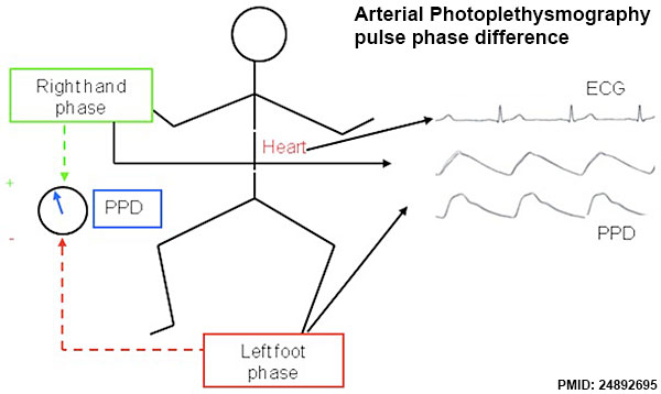
Photoplethysmography signals acquisition technique using infrared sensors
Acquired signals were used to compute pulse phases corresponding to preductal (right hand sensor) and postductal (foot sensor) regions and their difference, the PPD. doi:10.1371/journal.pone.0098763.g001
Reference
<pubmed>24892695</pubmed>| PLoS One.
Goudjil S, Imestouren F, Armougon A, Razafimanantsoa L, Mahmoudzadeh M, et al. (2014) Noninvasive Technique for the Diagnosis of Patent Ductus Arteriosus in Premature Infants by Analyzing Pulse Wave Phases on Photoplethysmography Signals Measured in the Right Hand and the Left Foot. PLoS ONE 9(6): e98763. doi:10.1371/journal.pone.0098763
Copyright
© 2014 Goudjil et al. This is an open-access article distributed under the terms of the Creative Commons Attribution License, which permits unrestricted use, distribution, and reproduction in any medium, provided the original author and source are credited.
File history
Click on a date/time to view the file as it appeared at that time.
| Date/Time | Thumbnail | Dimensions | User | Comment | |
|---|---|---|---|---|---|
| current | 23:23, 25 June 2014 | | 600 × 358 (31 KB) | Z8600021 (talk | contribs) | ===Reference=== <pubmed>24892695</pubmed>| [http://www.plosone.org/article/info%3Adoi%2F10.1371%2Fjournal.pone.0098763 PLoS One.] Goudjil S, Imestouren F, Armougon A, Razafimanantsoa L, Mahmoudzadeh M, et al. (2014) Noninvasive Technique for the Dia... |
You cannot overwrite this file.
File usage
The following page uses this file: