File:Odgers1939-plate01.jpg

Original file (1,200 × 1,314 pixels, file size: 519 KB, MIME type: image/jpeg)
Plate I
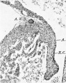
Fig. 1. A section through the right A.-V. orifice in an 11-2 mm. embryo ( x 109). It shows the right lateral cushion, 13.0., separated from the A.-V. sulcus, A. V.S., by auricular muscle, A., joining that of the ventricle, V.
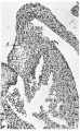
Fig. 2. A section of the right lateral cusp in a 17-5 mm. embryo ( x 109) to show the angulation of the right A.-V. sulcus, A. V.S., which is still separated from the right lateral cushion, R.0., by the junction of auricular, A., with ventricular muscle, V.
Fig. 3. A section through the left lateral cusp in the same embryo as fig. 2 ( x 109) to show in contrast the absence of any angulation of the sulcus, A.V.S., while‘ the left lateral cushion, L.0., maintains its original shape and relations to A., auricular muscle and to V., ventricular muscle.
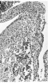
Fig. 4. A section through the heart in a 23 mm. embryo ( x 60). It shows the cushions of the central cusps, M .0., capping the muscular interventricular septum, I .V.S., and those of the lateral cusps, 12.0’. and L0. Note the angulation of the right A.-V. sulcus, A.V.S. A. auricular, V. ventricular muscle.
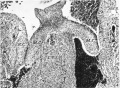
Fig. 5. A section through the right lateral cusp in a 28-5 mm. embryo ( x 60) to show the effect of the invagination which has now occurred at the right A.-V. sulcus, A. V.S. Auricular muscle, A., joins that of the ventricle, V., ventral to the line of attachment of the cusp, so that its base is now composed of auricular muscle, A., and ventricular muscle, 17., with a core of connective tissue continuous with that of the sulcus. The cushion tissue, R.C., extends almost to the tip of the cusp.
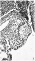
1 right A.-V. orifice in an 11-2 mm. embryo
2 right lateral cusp in a 17-5 mm. embryo
3 left lateral cusp in a 17-5 mm. embryo
4 central cusps in a 23 mm. embryo
5 right lateral cusp in a 28-5 mm. embryo


| Historic Disclaimer - information about historic embryology pages |
|---|
 |
Reference
Odgers PNB. The Development of the Atrio-Ventricular Valves in Man (1939) J Anat. 73:643-57. PMID 17104787
Cite this page: Hill, M.A. (2024, April 26) Embryology Odgers1939-plate01.jpg. Retrieved from https://embryology.med.unsw.edu.au/embryology/index.php/File:Odgers1939-plate01.jpg
- © Dr Mark Hill 2024, UNSW Embryology ISBN: 978 0 7334 2609 4 - UNSW CRICOS Provider Code No. 00098G
File history
Click on a date/time to view the file as it appeared at that time.
| Date/Time | Thumbnail | Dimensions | User | Comment | |
|---|---|---|---|---|---|
| current | 14:32, 15 November 2015 | | 1,200 × 1,314 (519 KB) | Z8600021 (talk | contribs) |
You cannot overwrite this file.
File usage
The following page uses this file: