File:Odgers1937 plate02fig02.jpg
From Embryology
Size of this preview: 800 × 554 pixels. Other resolution: 1,000 × 692 pixels.
Original file (1,000 × 692 pixels, file size: 157 KB, MIME type: image/jpeg)
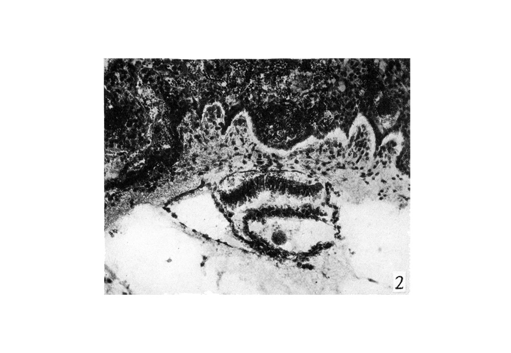
Fig. 2. A photograph of a section of the Peters ovum
Kindly sent me by Prof.0. Grosser. It shows the exocoelomic channel on the left intact, bordering the embryonic disc and the yolk sac, while that on the right has only its medial wall left.
(x100)
| Historic Disclaimer - information about historic embryology pages |
|---|
 |
Reference
Odgers PN. An early human ovum (Thomson) in situ. (1937) J Anat. 71(2): 161-168.3. PMID 17104634
Cite this page: Hill, M.A. (2024, April 26) Embryology Odgers1937 plate02fig02.jpg. Retrieved from https://embryology.med.unsw.edu.au/embryology/index.php/File:Odgers1937_plate02fig02.jpg
- © Dr Mark Hill 2024, UNSW Embryology ISBN: 978 0 7334 2609 4 - UNSW CRICOS Provider Code No. 00098G
File history
Click on a date/time to view the file as it appeared at that time.
| Date/Time | Thumbnail | Dimensions | User | Comment | |
|---|---|---|---|---|---|
| current | 21:20, 29 June 2015 | | 1,000 × 692 (157 KB) | Z8600021 (talk | contribs) |
You cannot overwrite this file.
File usage
The following page uses this file: