File:Odgers1937 plate01.jpg
Original file (1,446 × 2,000 pixels, file size: 919 KB, MIME type: image/jpeg)
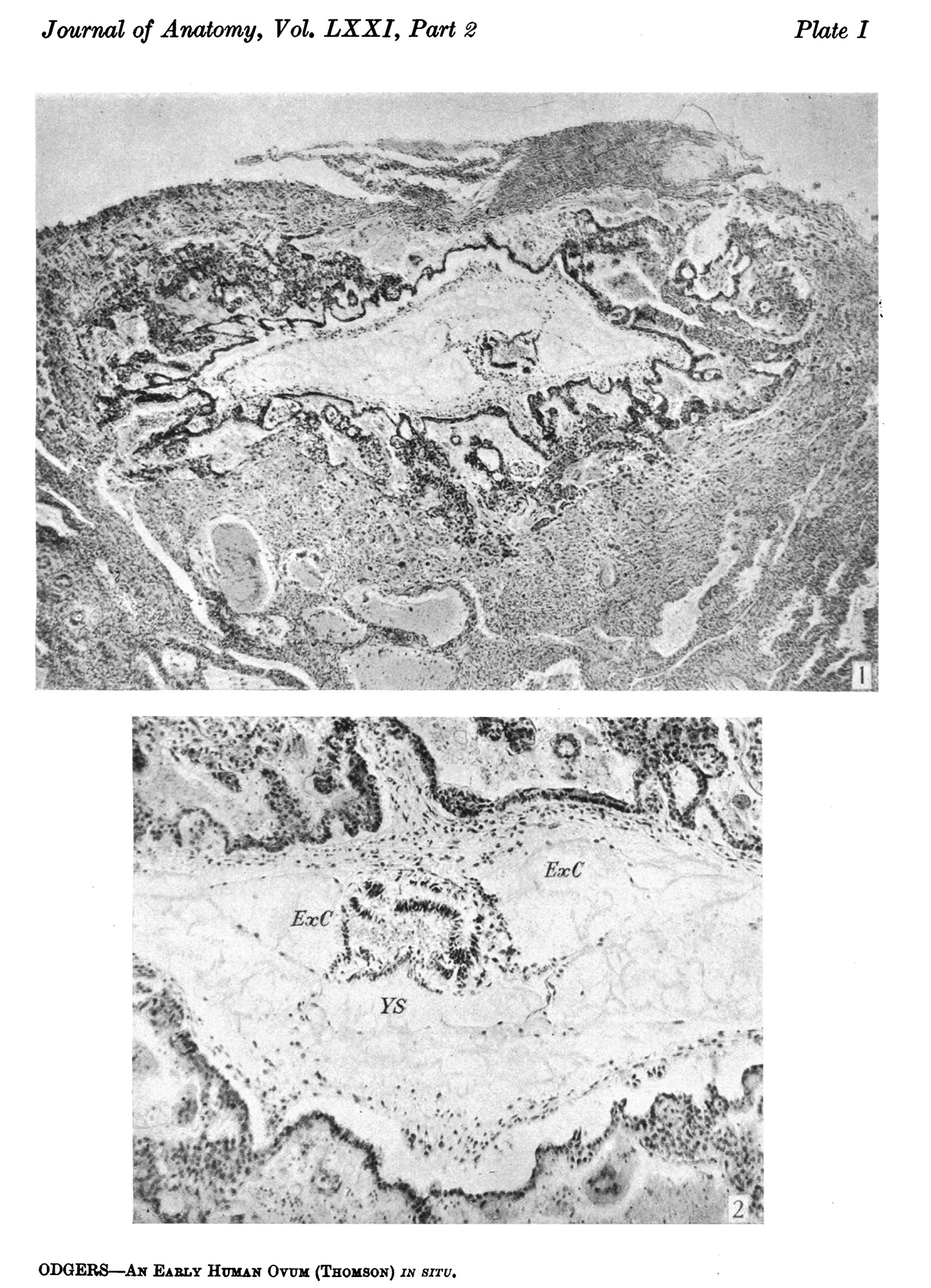
Plate 1
Fig. 1. The section photographed is through the middle of the blastocyst cavity (x44).
Fig. 2. The same section as fig. 1 (x 100). This shows the exocoelomic channels, ExC, on either side of the embryonic disc and ventral to this the yolk sac, YS. Above the disc is the amniotic cavity obscured by cellular debris which partially fills it. This section corresponds to the plane lettered B in Text-fig.1.
| Historic Disclaimer - information about historic embryology pages |
|---|
 |
Reference
Odgers PN. An early human ovum (Thomson) in situ. (1937) J Anat. 71(2): 161-168.3. PMID 17104634
Cite this page: Hill, M.A. (2024, April 26) Embryology Odgers1937 plate01.jpg. Retrieved from https://embryology.med.unsw.edu.au/embryology/index.php/File:Odgers1937_plate01.jpg
- © Dr Mark Hill 2024, UNSW Embryology ISBN: 978 0 7334 2609 4 - UNSW CRICOS Provider Code No. 00098G
File history
Click on a date/time to view the file as it appeared at that time.
| Date/Time | Thumbnail | Dimensions | User | Comment | |
|---|---|---|---|---|---|
| current | 21:03, 29 June 2015 | | 1,446 × 2,000 (919 KB) | Z8600021 (talk | contribs) | |
| 21:01, 29 June 2015 |  | 1,870 × 2,587 (1.46 MB) | Z8600021 (talk | contribs) |
You cannot overwrite this file.
File usage
The following page uses this file: