File:O'Connor1940 plate01.jpg

Original file (1,280 × 1,503 pixels, file size: 268 KB, MIME type: image/jpeg)
Plate I
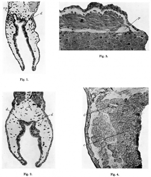
Fig. 1. Amblystoma, transverse section through the cloaca 15 days after operation to prevent the formation of the left pronephric duct. Symmetrical development of the cloaca and cloacal diverticula. (r.p. right pronephric duct; d, cloacal diverticulum.) x 120.
Fig. 2. Triton taentatus, transverse section through the cloaca 12 days after operation as indicated in Text-fig. 2. Normal development of the cloaca in the absence of both pronephric ducts. (d, cloacal diverticulum.) x 140.
Fig. 3. Triton taeniatus, longitudinal section 7 days after operation as described in text. The pronephric duct comes up against the ectoderm but does not unite with it. (p, pronephric duct.) x 120.
Fig. 4. Amblystoma, transverse section through the cloaca 40, cranial to the entrance of the pronephric ducts. 9 days previously embryo stained with Nile blue sulphate as indicated in Text-fig. 5. Nile blue sulphate seen as granules in the cloacal wall (c) and in the right pronephric duct (p). x 300.
Reference
O'Connor RJ. An experimental study of the development of the amphibian cloaca. (1940) J Anat. 74(3):301 - 308. PMID 17104815
Cite this page: Hill, M.A. (2024, April 26) Embryology O'Connor1940 plate01.jpg. Retrieved from https://embryology.med.unsw.edu.au/embryology/index.php/File:O%27Connor1940_plate01.jpg
- © Dr Mark Hill 2024, UNSW Embryology ISBN: 978 0 7334 2609 4 - UNSW CRICOS Provider Code No. 00098G
File history
Click on a date/time to view the file as it appeared at that time.
| Date/Time | Thumbnail | Dimensions | User | Comment | |
|---|---|---|---|---|---|
| current | 12:32, 14 November 2018 |  | 1,280 × 1,503 (268 KB) | Z8600021 (talk | contribs) | |
| 12:30, 14 November 2018 |  | 1,758 × 2,523 (755 KB) | Z8600021 (talk | contribs) | {{Ref-O'Connor1940}} |
You cannot overwrite this file.
File usage
There are no pages that use this file.