File:Muscle elongation.png
Muscle_elongation.png (518 × 600 pixels, file size: 194 KB, MIME type: image/png)
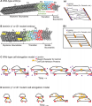
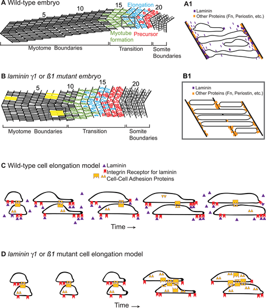
Cartoon of WT embryo showing the three phases of muscle elongation
In the oldest/most anterior somites, myotubes have formed and are attached to the MTJs. The transition region contains cells intercalating by extending protrusions that are subsequently filled. Muscle precursor cells exhibit protrusive activity in all directions. A1: Magnification of a somite in panel A showing proteins concentrated at the MTJ and boundary capture of recently elongated cells.
B–B1) Cartoon of laminin mutant embryo at the same age as WT embryo in panel A showing the same three phases of muscle morphogenesis, but with a developmental delay. Cells in yellow are aberrantly long and have invaded into neighboring myotomes. B1: Magnification of two somites in panel B that depicts a model of how boundary crossing could occur in laminin mutant embryos. If laminin is absent, there may be randomly spaced locations at the MTJ devoid of proteins that function in boundary capture. Elongating muscle cells would invade the MTJ at these locations.
C) Cartoon model showing the two-step mechanism of elongating. We show that adhesion to the matrix is required for normal elongation and hypothesize that cells also utilize cell-cell adhesion to generate traction forces needed for protrusion extension and filling.
D) Model accounting for developmental delay in muscle morphogenesis that occurs in laminin mutants. Cartoon depicting a laminin mutant cell undergoing two-step elongation via protrusion extension and filling. Lack of the cell-matrix adhesion protein laminin results in less traction and therefore slower extension and/or filling.
- "Skeletal muscle morphogenesis transforms short muscle precursor cells into long, multinucleate myotubes that anchor to tendons via the myotendinous junction (MTJ). In vertebrates, a great deal is known about muscle specification as well as how somitic cells, as a cohort, generate the early myotome. However, the cellular mechanisms that generate long muscle fibers from short cells and the molecular factors that limit elongation are unknown. We show that zebrafish fast muscle fiber morphogenesis consists of three discrete phases: short precursor cells, intercalation/elongation, and boundary capture/myotube formation. In the first phase, cells exhibit randomly directed protrusive activity. The second phase, intercalation/elongation, proceeds via a two-step process: protrusion extension and filling. This repetition of protrusion extension and filling continues until both the anterior and posterior ends of the muscle fiber reach the MTJ. Finally, both ends of the muscle fiber anchor to the MTJ (boundary capture) and undergo further morphogenetic changes as they adopt the stereotypical, cylindrical shape of myotubes. We find that the basement membrane protein laminin is required for efficient elongation, proper fiber orientation, and boundary capture. These early muscle defects in the absence of either lamininβ1 or lamininγ1 contrast with later dystrophic phenotypes in lamininα2 mutant embryos, indicating discrete roles for different laminin chains during early muscle development. Surprisingly, genetic mosaic analysis suggests that boundary capture is a cell-autonomous phenomenon. Taken together, our results define three phases of muscle fiber morphogenesis and show that the critical second phase of elongation proceeds by a repetitive process of protrusion extension and protrusion filling. Furthermore, we show that laminin is a novel and critical molecular cue mediating fiber orientation and limiting muscle cell length."
Reference
Citation: Snow CJ, Goody M, Kelly MW, Oster EC, Jones R, et al. (2008) Time-Lapse Analysis and Mathematical Characterization Elucidate Novel Mechanisms Underlying Muscle Morphogenesis. PLoS Genet 4(10): e1000219. doi:10.1371/journal.pgen.1000219
Journal.pgen.1000219.g008.png
http://www.plosgenetics.org/article/info%3Adoi%2F10.1371%2Fjournal.pgen.1000219
Copyright
© 2008 Snow et al.
This is an open-access article distributed under the terms of the Creative Commons Attribution License, which permits unrestricted use, distribution, and reproduction in any medium, provided the original author and source are credited.
Cite this page: Hill, M.A. (2024, April 26) Embryology Muscle elongation.png. Retrieved from https://embryology.med.unsw.edu.au/embryology/index.php/File:Muscle_elongation.png
- © Dr Mark Hill 2024, UNSW Embryology ISBN: 978 0 7334 2609 4 - UNSW CRICOS Provider Code No. 00098G
File history
Click on a date/time to view the file as it appeared at that time.
| Date/Time | Thumbnail | Dimensions | User | Comment | |
|---|---|---|---|---|---|
| current | 14:28, 11 September 2009 |  | 518 × 600 (194 KB) | S8600021 (talk | contribs) | Cartoon of WT embryo showing the three phases of muscle elongation. In the oldest/most anterior somites, myotubes have formed and are attached to the MTJs. The transition region contains cells intercalating by extending protrusions that are subsequently |
You cannot overwrite this file.
File usage
There are no pages that use this file.
