File:Mouse oogenesis 02.jpg
Original file (1,386 × 355 pixels, file size: 40 KB, MIME type: image/jpeg)
Mouse Oogenesis
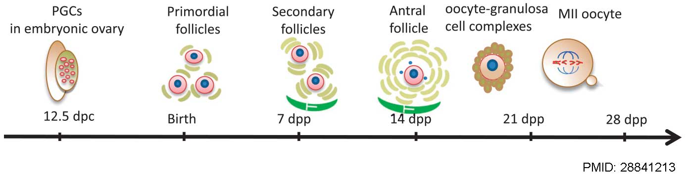
Schematic representation of the main stages of mouse oogenesis from 12.5 days post-coitum (dpc) and folliculogenesis, including the essential stages of embryonic primordial germ cells (PGCs) at 12.5 dpc, primordial follicles at birth, and the formation of secondary and antral follicles at 7 and 14 days post-partum (dpp), development of mature gametes as oocyte–granulosa cell complexes, oocyte meiosis and IVF.
Reference
Wang JJ, Ge W, Liu JC, Klinger FG, Dyce PW, De Felici M & Shen W. (2017). Complete in vitro oogenesis: retrospects and prospects. Cell Death Differ. , 24, 1845-1852. PMID: 28841213 DOI.
Copyright
This work is licensed under a Creative Commons Attribution 4.0 International License. The images or other third party material in this article are included in the article’s Creative Commons license, unless indicated otherwise in the credit line; if the material is not included under the Creative Commons license, users will need to obtain permission from the license holder to reproduce the material. To view a copy of this license, visit https://creativecommons.org/licenses/by/4.0/
Panel cropped from full figure 1 and PMID added.
Cite this page: Hill, M.A. (2024, April 26) Embryology Mouse oogenesis 02.jpg. Retrieved from https://embryology.med.unsw.edu.au/embryology/index.php/File:Mouse_oogenesis_02.jpg
- © Dr Mark Hill 2024, UNSW Embryology ISBN: 978 0 7334 2609 4 - UNSW CRICOS Provider Code No. 00098G
File history
Click on a date/time to view the file as it appeared at that time.
| Date/Time | Thumbnail | Dimensions | User | Comment | |
|---|---|---|---|---|---|
| current | 10:18, 8 March 2018 | 1,386 × 355 (40 KB) | Z8600021 (talk | contribs) |
You cannot overwrite this file.
File usage
The following page uses this file: