File:Mouse oogenesis 01.jpg

Original file (1,781 × 1,222 pixels, file size: 173 KB, MIME type: image/jpeg)
Mouse Oogenesis
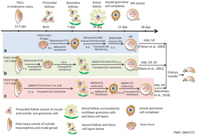
Schematic representation of the main stages of mouse oogenesis (upper drawing) and of the three in vitro culture methods of endogenous germ cells (medial drawings) capable of producing metaphase II (MII) oocytes that after in vitro maturation (IVM) and in vitro fertilization (IVF) or nuclear transfer (NT) generated live offspring; most essential components of the culture media are also reported.
The upper drawing displays the process of oogenesis from 12.5 days post-coitum (dpc) and folliculogenesis, including the essential stages of embryonic primordial germ cells (PGCs) at 12.5 dpc, primordial follicles at birth, and the formation of secondary and antral follicles at 7 and 14 days post-partum (dpp), development of mature gametes as oocyte–granulosa cell complexes, oocyte meiosis and IVF.
The medial figure shows the offspring through in vitro culture with 1 dpp ovary by O’Brien et al. (a) and fetal ovary at 12.5 dpc by Obata et al. (b) and Morohaku et al. (c) The lower panel shows the different cell types annotated (text from figure legend)
- Links: Oocyte Development | Ovary Development | Mouse Development
Reference
Wang JJ, Ge W, Liu JC, Klinger FG, Dyce PW, De Felici M & Shen W. (2017). Complete in vitro oogenesis: retrospects and prospects. Cell Death Differ. , 24, 1845-1852. PMID: 28841213 DOI.
Obata Y, Kono T & Hatada I. (2002). Gene silencing: maturation of mouse fetal germ cells in vitro. Nature , 418, 497. PMID: 12152066 DOI.
Morohaku K, Tanimoto R, Sasaki K, Kawahara-Miki R, Kono T, Hayashi K, Hirao Y & Obata Y. (2016). Complete in vitro generation of fertile oocytes from mouse primordial germ cells. Proc. Natl. Acad. Sci. U.S.A. , 113, 9021-6. PMID: 27457928 DOI.
Copyright
This work is licensed under a Creative Commons Attribution 4.0 International License. The images or other third party material in this article are included in the article’s Creative Commons license, unless indicated otherwise in the credit line; if the material is not included under the Creative Commons license, users will need to obtain permission from the license holder to reproduce the material. To view a copy of this license, visit https://creativecommons.org/licenses/by/4.0/
Cite this page: Hill, M.A. (2024, April 25) Embryology Mouse oogenesis 01.jpg. Retrieved from https://embryology.med.unsw.edu.au/embryology/index.php/File:Mouse_oogenesis_01.jpg
- © Dr Mark Hill 2024, UNSW Embryology ISBN: 978 0 7334 2609 4 - UNSW CRICOS Provider Code No. 00098G
File history
Click on a date/time to view the file as it appeared at that time.
| Date/Time | Thumbnail | Dimensions | User | Comment | |
|---|---|---|---|---|---|
| current | 10:11, 8 March 2018 | | 1,781 × 1,222 (173 KB) | Z8600021 (talk | contribs) | |
| 10:07, 8 March 2018 |  | 2,132 × 1,495 (287 KB) | Z8600021 (talk | contribs) | ==Schematic representation of the main stages of mouse oogenesis== (upper drawing) and of the three in vitro culture methods of endogenous germ cells (medial drawings) capable of producing metaphase II (MII) oocytes that after in vitro maturation (IVM... |
You cannot overwrite this file.
File usage
There are no pages that use this file.