File:Mouse oocyte and zona pellucida EM01.jpg
Original file (1,200 × 1,200 pixels, file size: 485 KB, MIME type: image/jpeg)
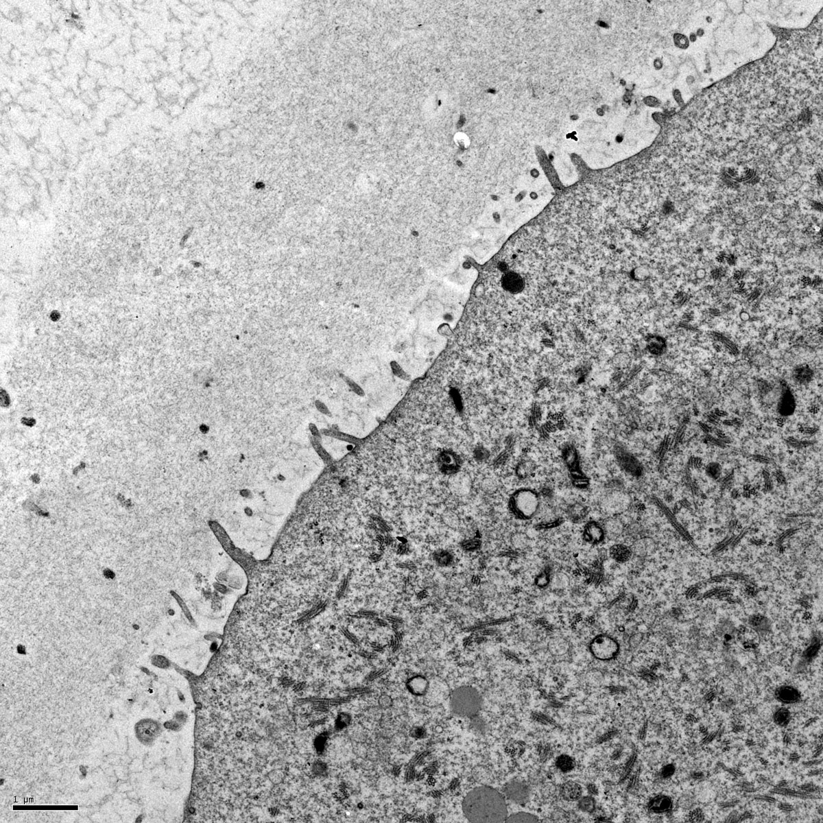
Mouse Oocyte Electron Micrograph
Surface of a mouse oocyte This transmission electron micrograph shows the cortex of a mouse oocyte that was undergoing fertilization. Microvilli are present on the oocyte's surface and the zona pellucida is visible surrounding the oocyte.
The scale bar is 1 micron (Stain - Osmium)
NCBI Organism Classification: Mus musculus
Cell Type: oocyte
Cellular Component: cell cortex, microvillus, zona pellucida
Related Images: large 1200px | 800px | Medium 600px | Small 400px
The sample was fixed using glutaraldehyde and osmium tetroxide, embedded in plastic, sectioned, and stained with uranyl acetate and lead citrate. The image was taken with a Phillips 500 transmission electron microscope.
Original Image: The Cell: An Image Library 12618.jpg http://www.cellimagelibrary.org/images/12618
Public Domain: This image is in the public domain and thus free of any copyright restrictions. However, as is the norm in scientific publishing and as a matter of courtesy, any user should credit the content provider for any public or private use of this image whenever possible.
File history
Click on a date/time to view the file as it appeared at that time.
| Date/Time | Thumbnail | Dimensions | User | Comment | |
|---|---|---|---|---|---|
| current | 07:58, 26 April 2011 | | 1,200 × 1,200 (485 KB) | S8600021 (talk | contribs) | ==Mouse Oocyte Electron Micrograph== Surface of a mouse oocyte This transmission electron micrograph shows the cortex of a mouse oocyte that was undergoing fertilization. The sample was fixed using glutaraldehyde and osmium tetroxide, embedded in plastic |
You cannot overwrite this file.
File usage
The following page uses this file: