File:Mouse oocyte Egr3-02.jpg
Original file (642 × 1,200 pixels, file size: 121 KB, MIME type: image/jpeg)
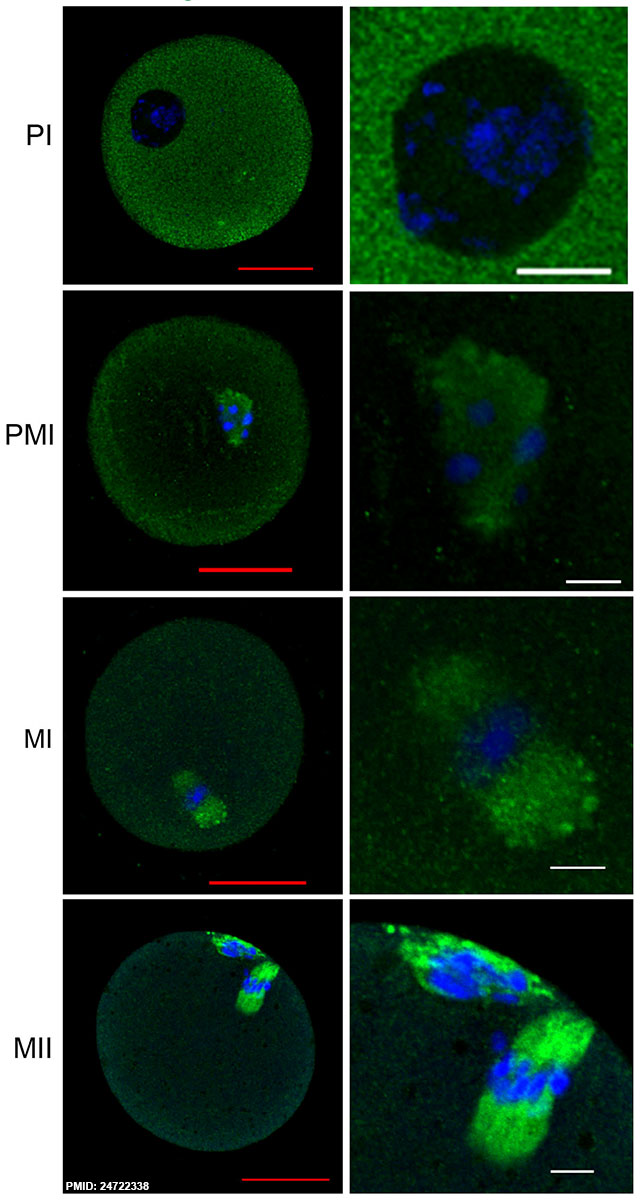
Mouse Oocyte Egr3 Expression
Egr3 is localized to the meiotic spindle of mouse oocytes at all stages of maturation. Germinal vesicle (GV) stage oocytes were obtained by ovary puncture and were cultured in M16. Oocytes were fixed in 3.7% formaldehyde and were subjected to immunofluorescence staining with anti-Egr3 antibody (Santa Cruz). Oocytes in the left panel are shown at 60X and enlarged images of chromosome-containing areas are shown in the right panel.
PI, prophase I; PMI, prometaphase I (cultured for 3 h); MI, metaphase I (cultured for 8 h); MII, metaphase II (cultured for 12 h).
Red scale bar, 30 μm; white scale bar, 10 μm. Green, Egr3; blue, DNA.
Reference
<pubmed>24722338</pubmed>| PLoS One.
Copyright
© 2014 Shin et al. This is an open-access article distributed under the terms of the Creative Commons Attribution License, which permits unrestricted use, distribution, and reproduction in any medium, provided the original author and source are credited.
Citation: Shin H, Kwon S, Song H, Lim HJ (2014) The Transcription Factor Egr3 Is a Putative Component of the Microtubule Organizing Center in Mouse Oocytes. PLoS ONE 9(4): e94708. doi:10.1371/journal.pone.0094708
Journal.pone.0094708.g001.jpg Fig 1 cropped, resized and relabeled.
File history
Click on a date/time to view the file as it appeared at that time.
| Date/Time | Thumbnail | Dimensions | User | Comment | |
|---|---|---|---|---|---|
| current | 22:45, 16 April 2014 | | 642 × 1,200 (121 KB) | Z8600021 (talk | contribs) | ==Mouse Oocyte Egr3 Expression== Egr3 localization in a mouse oocyte within a growing follicle. Meiotic spindles of maturing oocytes (Fig. 1A, arrow). Immunofluorescence staining of Egr3 was performed on ovarian cryosection fixed in acetone. The rab... |
You cannot overwrite this file.
File usage
There are no pages that use this file.