File:Mouse blastocyst trophoblast 01.jpg
Mouse_blastocyst_trophoblast_01.jpg (500 × 475 pixels, file size: 22 KB, MIME type: image/jpeg)
Localization of Trophoblast Marker
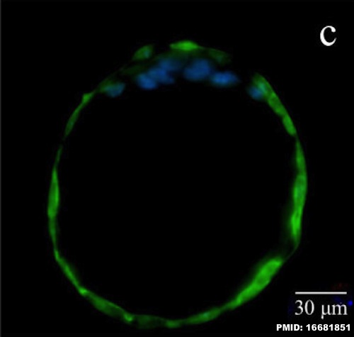
Hatched mouse blastocyst labelled for a marker for trophectoderm specification (TROMA-1). TROMA-1 antibody against cytokeratin 8, Endo A a cytokeratin-like filaments present in trophectoderm and endodermal cells.
- green - TROMA-1 expression
- blue - DAPI was used for nuclear DNA counterstaining.
Scale = 30 μm.
- Links: Blastocyst | Week 1 | Trophoblast
Reference
<pubmed>16681851</pubmed>| PMC1479373 | J Transl Med.
Copyright
© 2006 Tanaka et al; licensee BioMed Central Ltd. This is an Open Access article distributed under the terms of the Creative Commons Attribution License (http://creativecommons.org/licenses/by/2.0), which permits unrestricted use, distribution, and reproduction in any medium, provided the original work is properly cited.
Figure 5. (1479-5876-4-20-5-l.jpg) panel c cropped from full image, resized and relabelled.
Cite this page: Hill, M.A. (2024, April 25) Embryology Mouse blastocyst trophoblast 01.jpg. Retrieved from https://embryology.med.unsw.edu.au/embryology/index.php/File:Mouse_blastocyst_trophoblast_01.jpg
- © Dr Mark Hill 2024, UNSW Embryology ISBN: 978 0 7334 2609 4 - UNSW CRICOS Provider Code No. 00098G
File history
Click on a date/time to view the file as it appeared at that time.
| Date/Time | Thumbnail | Dimensions | User | Comment | |
|---|---|---|---|---|---|
| current | 11:56, 5 May 2013 |  | 500 × 475 (22 KB) | Z8600021 (talk | contribs) | ==Localization of Trophoblast Marker== Hatched mouse blastocyst labelled for a marker for trophectoderm specification (TROMA-1). TROMA-1 antibody against cytokeratin 8, Endo A a cytokeratin-like filaments present in trophectoderm and endodermal cells.... |
You cannot overwrite this file.
File usage
The following page uses this file:
