File:Mouse antral follicle.jpg

Original file (600 × 705 pixels, file size: 168 KB, MIME type: image/jpeg)
Antral Follicle
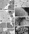
FSH antral follicles: oocyte and granulosa cells. The oocytes (O) contain numerous organelles, uniformly distributed in the ooplasm (panels a, b, d). Note the presence of scattered cortical granules (arrows) in the suboolemmal area (panel b). Numerous microvilli (m) can be observed on the oocyte surface (panels b, c). Cytoplasmic projections (arrows) stemming from the inner granulosa cell (GC) layer are seen crossing the zona pellucida (ZP) (panels a, d) and reaching the oocyte (transzonal processes) (panels d, e). Areas of detachment between inner granulosa cells and oocyte are also present (asterisks, panels a, d). Outer granulosa cells (GC) appeared irregularly rounded/polygonal and scarcely adherent each other (panels f, h). Uniformly dispersed chromatin and one or more nucleoli are seen in both inner (panels a, d) and outer (panels f, g) granulosa cells. Panel b, ZP: zona pellucida. Panel e, O: oocyte. Panel f, arrow: loss of contact among granulosa cells; panel g, L: lipid droplets in the granulosa cell cytoplasm. Bar is: 3.5 μm (panel a); 1 μm (panel b); 10 μm (panel c); 2 μm (panel d); 9 μm (panel e); 3 μm (panels f, g); 50 μm (panel h). Panels a, b, d, f, g: TEM; panels c, e, h: SEM.
1477-7827-9-3-4.jpg
Reference
<pubmed>21232101</pubmed>Reprod Biol Endocrinol.
© 2011 Nottola et al; licensee BioMed Central Ltd. This is an Open Access article distributed under the terms of the Creative Commons Attribution License (http://creativecommons.org/licenses/by/2.0), which permits unrestricted use, distribution, and reproduction in any medium, provided the original work is properly cited.
Nottola et al. Reproductive Biology and Endocrinology 2011 9:3 doi:10.1186/1477-7827-9-3
File history
Click on a date/time to view the file as it appeared at that time.
| Date/Time | Thumbnail | Dimensions | User | Comment | |
|---|---|---|---|---|---|
| current | 23:44, 23 February 2011 |  | 600 × 705 (168 KB) | S8600021 (talk | contribs) | ==Antral Follicle== FSH antral follicles: oocyte and granulosa cells. The oocytes (O) contain numerous organelles, uniformly distributed in the ooplasm (panels a, b, d). Note the presence of scattered cortical granules (arrows) in the suboolemmal area (p |
You cannot overwrite this file.
File usage
There are no pages that use this file.