File:Mouse- postnatal osteoblasts.jpg
Mouse-_postnatal_osteoblasts.jpg (400 × 537 pixels, file size: 59 KB, MIME type: image/jpeg)
Mouse Osteoblasts
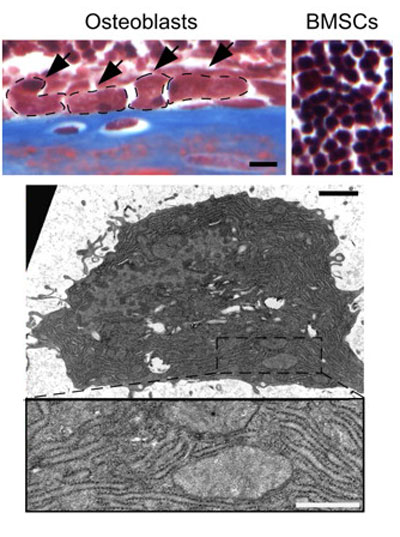
Masson’s trichrome staining of postnatal day 10 (P10) tibial sections. Arrows indicate osteoblasts lining the endosteal bone surface and osteoblasts are delineated by the dashed lines. Bone marrow stromal cells (BMSCs; right) Bar, 10 µm.
Transmission electron microscopy of osteoblasts in P10 tibiae.
Higher magnification views of boxed regions show extensive, well-organized rER cisternae. Bars: (top) 1 µm; (bottom) 0.5 µm.
Original Image File: Figure 7. Opt is essential for type I collagen synthesis in differentiating osteoblasts. http://jcb.rupress.org/content/189/3/511/F7.expansion.html (Image extracted from full figure)
Reference
<pubmed>20440000</pubmed>| JCB
Sohaskey M L et al. J Cell Biol 2010;189:511-525
Copyright
Rockefeller University Press - Copyright Policy This article is distributed under the terms of an Attribution–Noncommercial–Share Alike–No Mirror Sites license for the first six months after the publication date (see http://www.jcb.org/misc/terms.shtml). After six months it is available under a Creative Commons License (Attribution–Noncommercial–Share Alike 4.0 Unported license, as described at https://creativecommons.org/licenses/by-nc-sa/4.0/ ). (More? Help:Copyright Tutorial)
File history
Click on a date/time to view the file as it appeared at that time.
| Date/Time | Thumbnail | Dimensions | User | Comment | |
|---|---|---|---|---|---|
| current | 12:16, 24 November 2010 |  | 400 × 537 (59 KB) | S8600021 (talk | contribs) | ==Mouse Osteoblasts== Masson’s trichrome staining of postnatal day 10 (P10) tibial sections. Arrows indicate osteoblasts lining the endosteal bone surface and osteoblasts are delineated by the dashed lines. Bone marrow stromal cells (BMSCs; right) Bar, |
You cannot overwrite this file.
File usage
There are no pages that use this file.
