File:Morton1949 fig03.jpg
From Embryology
Size of this preview: 775 × 600 pixels. Other resolution: 1,016 × 786 pixels.
Original file (1,016 × 786 pixels, file size: 108 KB, MIME type: image/jpeg)
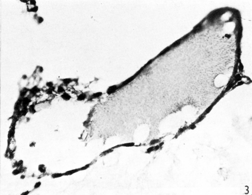
Fig. 3. The distal expansion of the yolk-sac
The end of the duct just appears above, on the left. Sect.77. x375.
| Historic Disclaimer - information about historic embryology pages |
|---|
 |
- Links: Text-fig. 1 | Text-fig. 2 | Fig. 1 | Fig. 2 | Fig. 3 | Fig. 4 | Fig. 5 | Fig. 6 | Plate 1 | Plate 2
Reference
Morton WRM. Two early human embryos. (1949) J. Anat., 83: 308-314.
Cite this page: Hill, M.A. (2024, April 26) Embryology Morton1949 fig03.jpg. Retrieved from https://embryology.med.unsw.edu.au/embryology/index.php/File:Morton1949_fig03.jpg
- © Dr Mark Hill 2024, UNSW Embryology ISBN: 978 0 7334 2609 4 - UNSW CRICOS Provider Code No. 00098G]
File history
Click on a date/time to view the file as it appeared at that time.
| Date/Time | Thumbnail | Dimensions | User | Comment | |
|---|---|---|---|---|---|
| current | 19:55, 11 August 2015 | | 1,016 × 786 (108 KB) | Z8600021 (talk | contribs) |
You cannot overwrite this file.
File usage
The following 2 pages use this file: