File:Model mifepristone bound to progesterone receptor.jpg
Original file (1,280 × 514 pixels, file size: 171 KB, MIME type: image/jpeg)
Model mifepristone bound to progesterone receptor
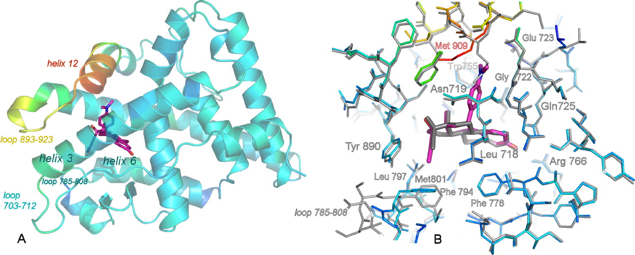
Binding of mifepristone (RU486, purple) in the progesterone receptor ligand binding domain (PR LBD, blue/red).
A - ribbon view of the PR LBD copy in the dimer that binds RU486. The coloring represents B-factor changes compared with the equiconformational LBD copy in the PR norethindrone complex. Changes range from −4 (blue) to +44 (red) Å2 and are predominantly restricted to the loop 785–808 and helix 12. The conformation of helix 12 is agonistic, closely packed against the LBD core.
B - superposition of the two PR LBD copies in the dimer, one containing RU486 (coloring as in panel A) and one containing norethindrone (gray). Important residues and loops are marked.
- Links: Mifepristone
Reference
<pubmed>19372222</pubmed>
Copyright
Copyright © 2009, by the American Society for Biochemistry and Molecular Biology
Non-profit/Non-commercial Use
For Non-profit/Non-commercial uses: You are free to copy, distribute, transmit and to adapt the work under the following conditions:
Attribution. You must attribute the work in the manner specified by the author or licensor (but not in any way that suggests that they endorse you or your use of the work).
Non-commercial. You may not use the work for commercial purposes; including original authors reusing content by a commercial publisher.
File history
Click on a date/time to view the file as it appeared at that time.
| Date/Time | Thumbnail | Dimensions | User | Comment | |
|---|---|---|---|---|---|
| current | 12:56, 5 May 2013 | 1,280 × 514 (171 KB) | Z8600021 (talk | contribs) | Binding of mifepristone (RU486, purple) in the progesterone receptor ligand binding domain (PR LBD, blue/red). A - ribbon view of the PR LBD copy in the dimer that binds RU486. The coloring represents B-factor changes compared with the equiconformatio... |
You cannot overwrite this file.
File usage
The following page uses this file: