File:Meyer1920 fig19.jpg
Meyer1920_fig19.jpg (575 × 389 pixels, file size: 33 KB, MIME type: image/jpeg)
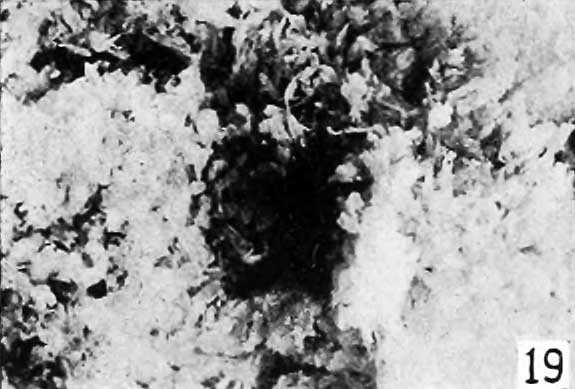
Fig. 19.
Small chorionic vesicles, such as No. 2077 shown in natural size in figure 18, which attract no attention upon cursory inspection may, and often do, present the most exquisite picture of hydatiform degeneration when seen under a magnification of 3 to 20 diameters, as illustrated in figure 19. This is true especially if the examination is made with the binocular microscope. Since I have adopted this method of examination it has been possible to recognize instances of decidedly general and typical hydatiform degeneration in chorionic vesicles less than 2 cm. in size, with later confirmation of the diagnosis by a histologic examination. However, I have not been able to recognize very early stages merely by examination of the gross specimens, for gross recognition is possible only when portions of at least some of the villi have become sufficiently elliptical or globular to attract attention. Histologic recognition is possible far earlier than this, as shown in figure 20.
- Meyer Links: Plate 1 | Plate 2 | Plate 3 | Plate 4 | Plate 5 | Plate 6 | Contribution No.40 | Volume IX | Contributions to Embryology | Hydatidiform Mole | Tubal Pregnancy
| Historic Disclaimer - information about historic embryology pages |
|---|
 |
Reference
Meyer AW. Hydatiform degeneration in tubal and uterine pregnancy. (1920) Carnegie Instn. Wash. Publ., Contrib. Embryol., 40: 327- 364.
Cite this page: Hill, M.A. (2024, April 26) Embryology Meyer1920 fig19.jpg. Retrieved from https://embryology.med.unsw.edu.au/embryology/index.php/File:Meyer1920_fig19.jpg
- © Dr Mark Hill 2024, UNSW Embryology ISBN: 978 0 7334 2609 4 - UNSW CRICOS Provider Code No. 00098G
File history
Click on a date/time to view the file as it appeared at that time.
| Date/Time | Thumbnail | Dimensions | User | Comment | |
|---|---|---|---|---|---|
| current | 10:33, 8 April 2012 | | 575 × 389 (33 KB) | Z8600021 (talk | contribs) | ==Fig. 19.== Plate 3: Fig. 14 | Fig. 15 | Fig. 16 | Fig. 17 | Fig. 18 | |
You cannot overwrite this file.
File usage
The following page uses this file: