File:Meyer1914 fig01-02.jpg
From Embryology

Size of this preview: 800 × 442 pixels. Other resolution: 1,266 × 700 pixels.
Original file (1,266 × 700 pixels, file size: 110 KB, MIME type: image/jpeg)
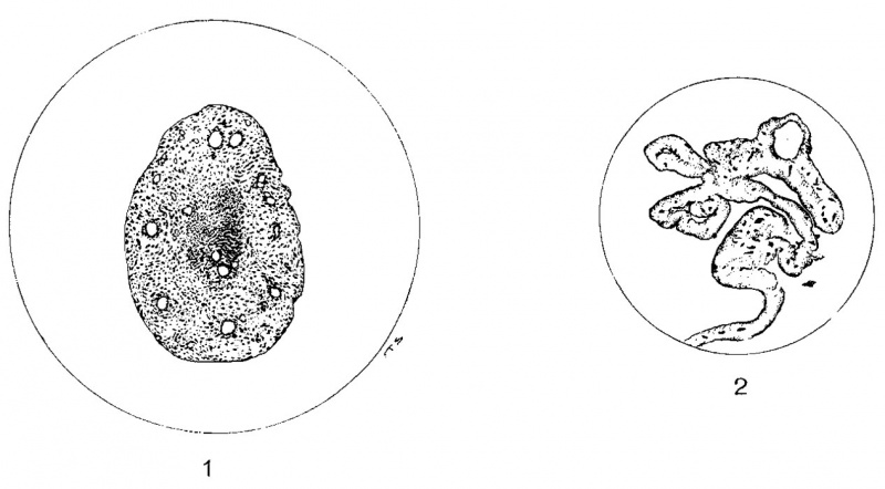
Fig. 1 Degenerating umbilical vein of a lamb 91 hours old. X142.
Fig. 2 Plicated caudal border of the suspensory ligament of a rabbit. In some portions these folds are fused. X275.
| Historic Disclaimer - information about historic embryology pages |
|---|
 |
Reference
Cite this page: Hill, M.A. (2024, April 26) Embryology Meyer1914 fig01-02.jpg. Retrieved from https://embryology.med.unsw.edu.au/embryology/index.php/File:Meyer1914_fig01-02.jpg
- © Dr Mark Hill 2024, UNSW Embryology ISBN: 978 0 7334 2609 4 - UNSW CRICOS Provider Code No. 00098G
File history
Click on a date/time to view the file as it appeared at that time.
| Date/Time | Thumbnail | Dimensions | User | Comment | |
|---|---|---|---|---|---|
| current | 21:02, 3 November 2015 | | 1,266 × 700 (110 KB) | Z8600021 (talk | contribs) | |
| 21:02, 3 November 2015 |  | 1,318 × 847 (165 KB) | Z8600021 (talk | contribs) | Fig. 1 Degenerating umbilical vein of a lamb 91 hours old. X142. Fig. 2 Plicated caudal border of the suspensory ligament of a rabbit. In some portions these folds are fused. X275. |
You cannot overwrite this file.
File usage
The following page uses this file: