File:MartinFalkiner1938 fig02.jpg

Original file (600 × 886 pixels, file size: 76 KB, MIME type: image/jpeg)
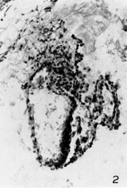
Fig 2
Section 29 A. X 150. The large amniotic cavity lies dorsal and it is prolonged into the body stalk as the amniotic duct. The small cavity of the yolk sac is separated from the amnion by a considera.ble interval filled with loose tissue with deep grooves on either side. In the floor of the amniotic cavity the extreme edge of the embryonic p-late has been sectioned very obliquely. The allantois, though indistinct, can be seen in the body stalk.
Reference
Martin CP. and Falkiner N. Mcl. The Falkiner ovum. (1938) Amer. J Anat., 63: 251-271.
Cite this page: Hill, M.A. (2024, April 27) Embryology MartinFalkiner1938 fig02.jpg. Retrieved from https://embryology.med.unsw.edu.au/embryology/index.php/File:MartinFalkiner1938_fig02.jpg
- © Dr Mark Hill 2024, UNSW Embryology ISBN: 978 0 7334 2609 4 - UNSW CRICOS Provider Code No. 00098G
File history
Click on a date/time to view the file as it appeared at that time.
| Date/Time | Thumbnail | Dimensions | User | Comment | |
|---|---|---|---|---|---|
| current | 11:46, 11 August 2017 |  | 600 × 886 (76 KB) | Z8600021 (talk | contribs) |
You cannot overwrite this file.
File usage
The following 3 pages use this file: