File:Mall1916 plate01.jpg

Original file (1,298 × 1,500 pixels, file size: 359 KB, MIME type: image/jpeg)
Plate 1. Human Embryos - Abnormal
Fig. 1, 2. Photographs of the two halves of ovum No. 604. Natural size. The cavity of the ovum is filled with a jelly-like substance in which a pathological embryo is embedded.
Fig. 3. Section through a normal ovum No. 836, encapsulated in the decidua. X3. Drawn by Mr. Didusch. The embryo lies within the coelom, and bands of -magma fibrils radiate from the amnion to the chorionic wall. The head of the embryo shines through the more transparent portion of the amnion.
Fig. 4. Photograph of a block of the ovum, No. 536, in situ after the embryo had been removed. X25. The suporting strands of magma are strikingly shown.
Fig. 5. Pathological embryo No. 1117, embedded in hyaline magma. X4. From a tubal pregnancy following gonorrhea (?).
Fig. 6. Pathological ovum No. 560, containing a great quantity of reticular magma. X2i. The embryo is normal in form. From a case of retroversion of the uterus.
Fig. 7. Pathological ovum No. 991, with the cavity completely filled with reticular magma. Natural size. The embryo is normal inform. From a negro woman. Sect ions of the embryo indicate that it is macerated.
Fig. 8. Pathological ovum No. 531, containing a granular deposit around a nodular embryo. X 1 .
Fig. 9. Pathological ovum with a nodular embryo (651?). X2. The exocoelom is gorged with granular magma.
Fig. 10. Specimen No. 636. X. The embryo and chorion are normal in form, but the reticular magma is markedly increased in quantity.
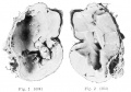
Fig. 1+2
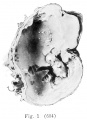
Fig. 1
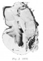
Fig. 2
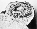
Fig. 3
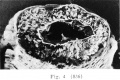
Fig. 4
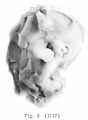
Fig. 5
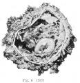
Fig. 6
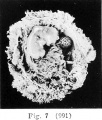
Fig. 7
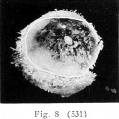
Fig. 8
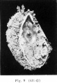
Fig. 9
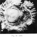
Fig. 10


| Historic Disclaimer - information about historic embryology pages |
|---|
 |
- Mall 1916 Links: Table 1 Normal Embryos | Table 2 Abnormal Embryos |Plate 1 | Fig. 1+2 | Fig. 1 | Fig. 2 | Fig. 3 | Fig. 4 | Fig. 5 | Fig. 6 | Fig. 7 | Fig. 8 | Fig. 9 | Fig. 10 | Plate 2 | Fig. 1 | Fig. 2 | Fig. 3 | Plate 3 | Fig. 1 | Fig. 2 | Fig. 3 | Fig. 4+5 | Fig. 4 | Fig. 5 | Contributions to Embryology No.10
File history
Click on a date/time to view the file as it appeared at that time.
| Date/Time | Thumbnail | Dimensions | User | Comment | |
|---|---|---|---|---|---|
| current | 10:37, 22 April 2014 | | 1,298 × 1,500 (359 KB) | Z8600021 (talk | contribs) | ==Plate 1. == {{Mall1916 figures}} |
You cannot overwrite this file.
File usage
The following page uses this file: