File:Long1911 plate03.jpg

Original file (2,335 × 3,271 pixels, file size: 370 KB, MIME type: image/jpeg)
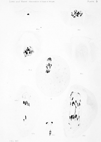
Plate 3 Division of First Spindle and Abstriction of First Polar Cell
(Figures 14 to 18, Inclusive).
Fig. 14. Ovarian egg containing an oblique spindle. Several of the chromosomes have already divided. Circumpolar bodies numerous and conspicuous. X (1200)960.
Fig. 14a. One chromosome from the spindle in fig. 14.
Figs. 15a, 156. An oblique spindle in two consecutive sections, showing the migration of the daughter chromosomes. Ovarian egg. X (2500) 2000.
Figs. 16a-16d. Four consecutive sections of a spindle similar in stage of division to that of fig. 17. See fig. H (p. 34). Ovarian egg. X(25oo) 2000.
| Historic Disclaimer - information about historic embryology pages |
|---|
| Pages where the terms "Historic" (textbooks, papers, people, recommendations) appear on this site, and sections within pages where this disclaimer appears, indicate that the content and scientific understanding are specific to the time of publication. This means that while some scientific descriptions are still accurate, the terminology and interpretation of the developmental mechanisms reflect the understanding at the time of original publication and those of the preceding periods, these terms, interpretations and recommendations may not reflect our current scientific understanding. (More? Embryology History | Historic Embryology Papers) |
File history
Click on a date/time to view the file as it appeared at that time.
| Date/Time | Thumbnail | Dimensions | User | Comment | |
|---|---|---|---|---|---|
| current | 20:22, 21 April 2014 |  | 2,335 × 3,271 (370 KB) | Z8600021 (talk | contribs) | ==Plate 3== {{Long1911 figures}} |
You cannot overwrite this file.
File usage
The following page uses this file:
