File:Long1911 plate01.jpg
Original file (1,079 × 1,500 pixels, file size: 95 KB, MIME type: image/jpeg)
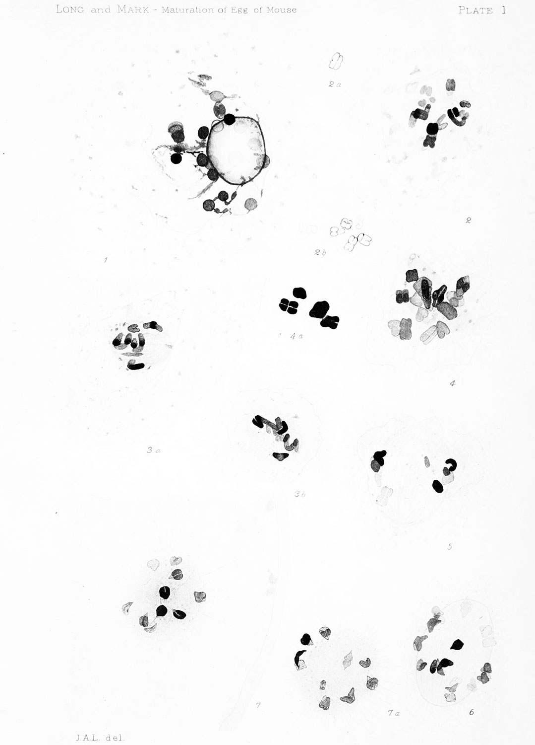
Plate 1 Origin of First Maturation Spindle
Fig. 1 . Germinative vesicle shortly before the disappearance of its nucleolus and the transformation of its contents into the fundaments of the chromosomes and the spindle fibers. Ovarian egg. X (2500) 2000.
Fig. 2. Early stage in the formation of the chromosome fundaments. Ovarian egg. X(25oo) 2000.
Figs. 2a, 2b. Fundaments of chromosomes in sections adjacent to that of fig. 2.
Figs. 3,a, 3b. Two consecutive sections showing a somewhat later stage than the preceding. Ovarian egg. X (2500) 2000.
Figs. 4, 4a. Chromosomes (20 in number) more completely differentiated. Spindle not yet formed. Nuclear membrane still intact. Ovarian egg. X (2500)2000.
Fig. 5. Section of a young spindle showing faint, fibrillations. There are 20 chromosomes scattered over its surface. Nuclear membrane is dissolved at some points. Ovarian egg. X (2500)2000.
Fig. 6. Composite drawing of a spindle cut into three parts. There are 20 chromosomes. Stage slightly more advanced than that illustrated in fig. 5. Nuclear membrane completely vanished. Ovarian egg. X (2500)2000.
Fig. 7. Two consecutive sections of a spindle, like that shown in fig. 6, seen in end view. There are 20 chromosomes, 10 in each section. The cytoplasm shows faint radiations about the spindle. Ovarian egg. X (2500)2000.
| Historic Disclaimer - information about historic embryology pages |
|---|
 |
File history
Click on a date/time to view the file as it appeared at that time.
| Date/Time | Thumbnail | Dimensions | User | Comment | |
|---|---|---|---|---|---|
| current | 20:21, 21 April 2014 | | 1,079 × 1,500 (95 KB) | Z8600021 (talk | contribs) | ==Plate 1== {{Long1911 figures}} |
You cannot overwrite this file.
File usage
The following page uses this file: