File:Lockwood1887b plate02.jpg

Original file (2,998 × 2,272 pixels, file size: 1.21 MB, MIME type: image/jpeg)
Plate II
Fig. 23. Glomerulus of human Wolffian body. Gl, glomerulus ; 7, Wolffian tubule; V, afferent and efferent vessels. 7 Hartnack. 4 Eye p.
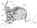
Fig. 24. Glomerulus of human Wolffian body, seventh week, showing the commencement of the development of capillaries in the glomerulus, the formation of the parenchyma and the tubules. P, parenchyma; t, tubule; Gi, glomerulus; C, capillaries ; e, epithelium of a commencing tubule. 7 Hartnack. 4 Eye p.
Fig. 25. Glomerulus of human Wolffian body at tenth week, showing vascularity of glomerulus and stroma of Wolffian body. Pr, parenchyma; Gi, glomerulus; WZ, Wolffian tubule. 7 Hartnack. 4 Kye p.
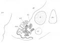
Fig. 26. Sexual eminence of rabbit, ,4, in oil immersion, to show relation of surface epithelium to meshes of stroma. This drawing was from a section close to that shown in fig. 37, p. 48.
Fig. 27. Urogenital ridge of rabbit, beginning of thirteenth day. M, mesentery; GH, germinal epithelium; WZ, Wolffian tubule ; WD, Wolffian duct; AO, aorta. 7 Hartnack. 4 Eye p.
Fig. 28. Human embryo, sexual eminence. GH, germinal epithelium ; CV, cardinal vein; WD, Wolffian duct; Gi, glomerulus. The bulk of the urogenital ridge and its genital eminence consists of mesoblastic cells of various shapes—round, branched, and elongated ; these have not been delineated. 7 Hartnack. 4 Eye p.
Fig. 29. Kidney and hinder part of the Wolffian body of a rabbit of thirteenth day. AB, kidney blastema; U, ureter; LCT, loose tissue, which surrounds kidney blastema ; WD, Wolffian duct; W7;, Wolffian tubules; PC, peritoneal cavity. x 70.
Fig. 30. Rabbit, fourteenth day, to show relation of hinder part of the Wolffian body and genital mass to one another, and to the kidney which has just appeared. GJ, genital mass; AO, aorta; XK, kidney ; HZ, hind limb; M, mesentery. x 25.
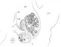
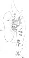
Fig. 31. Human embryo, thirty-five days. GW, genital mass ; K, kidney, lower end; AO, aorta; CV, cardinal vein; WZ, Wolffian tubules : ; Gl, glomerulus. x 45.
Fig. 32. Genital mass of rabbit, commencement of fourteenth day, to show stroma of branched anastomosing cells, and large, pale, granular cells in its meshes, 7 Hartnack. 4 Eye p.
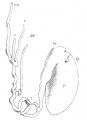
Fig. 33. Testicle and epididymis of human embryo, at about the twelfth week of intrauterine life. The section is not quite longitudinal. x 25. SZ, seminal tubules; RT, rete testes; Mesor., mesorchium ; Vas. Def., vasa deferens; VE, vasa efferentia; I7, indifferent tissue ; 7A, tunica albuginea.
Fig. 34. Outermost and front part of the same human Wolffian body as that which has been drawn in fig. 39, p. 61. WD, Wolffian duct; C7, collecting tube; ZZ, tubuli efferentia; GM, genital mass. x 45.
Fig. 35. Human foetus, eight months, to show the cell strings of the mediastinum testes which unite tubules of epididymis to seminiferous tubules. TE, tubules of epididymis; MT, mediastinum testes; ST, seminal tubules; V, blood-vessels. x 45.
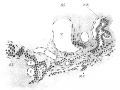
Fig. 36. Human testicle showing four vasa aberrantia. T, testicle; Hy, hydatid of Morgagni; VD, vas deferens; Hp, epididymis ; 1, 2, 3, and 4, vasa aberrantia.
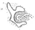
23 Glomerulus of human Wolffian body
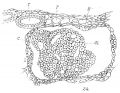
24 Glomerulus of human Wolffian body 7th week
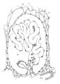
25 Glomerulus of human Wolffian body at 10th week
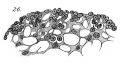
26 Sexual eminence of rabbit,
27 Urogenital ridge rabbit 13th day
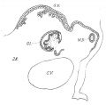
28 Human embryo, sexual eminence
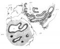
29 Kidney and part of the Wolffian body rabbit at 13th day
30 Rabbit 14th day hinder part of the Wolffian body and genital mass
31 Human embryo 35 days
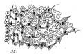
32 Genital mass rabbit at 14th day
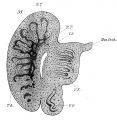
33 Testicle and epididymis of human embryo at about 12th week
34 Outermost and front part of the human Wolffian body
35 Human foetus 8th months
36 Human testicle showing four vasa aberrantia


Reference
Lockwood CB. Development and transition of the testis, normal and abnormal. (1887) J Anat. 22(1): 38-77. PMID 17231729
Cite this page: Hill, M.A. (2024, April 26) Embryology Lockwood1887b plate02.jpg. Retrieved from https://embryology.med.unsw.edu.au/embryology/index.php/File:Lockwood1887b_plate02.jpg
- © Dr Mark Hill 2024, UNSW Embryology ISBN: 978 0 7334 2609 4 - UNSW CRICOS Provider Code No. 00098G
File history
Click on a date/time to view the file as it appeared at that time.
| Date/Time | Thumbnail | Dimensions | User | Comment | |
|---|---|---|---|---|---|
| current | 22:02, 13 April 2020 | | 2,998 × 2,272 (1.21 MB) | Z8600021 (talk | contribs) | contrast |
| 18:50, 13 April 2020 | 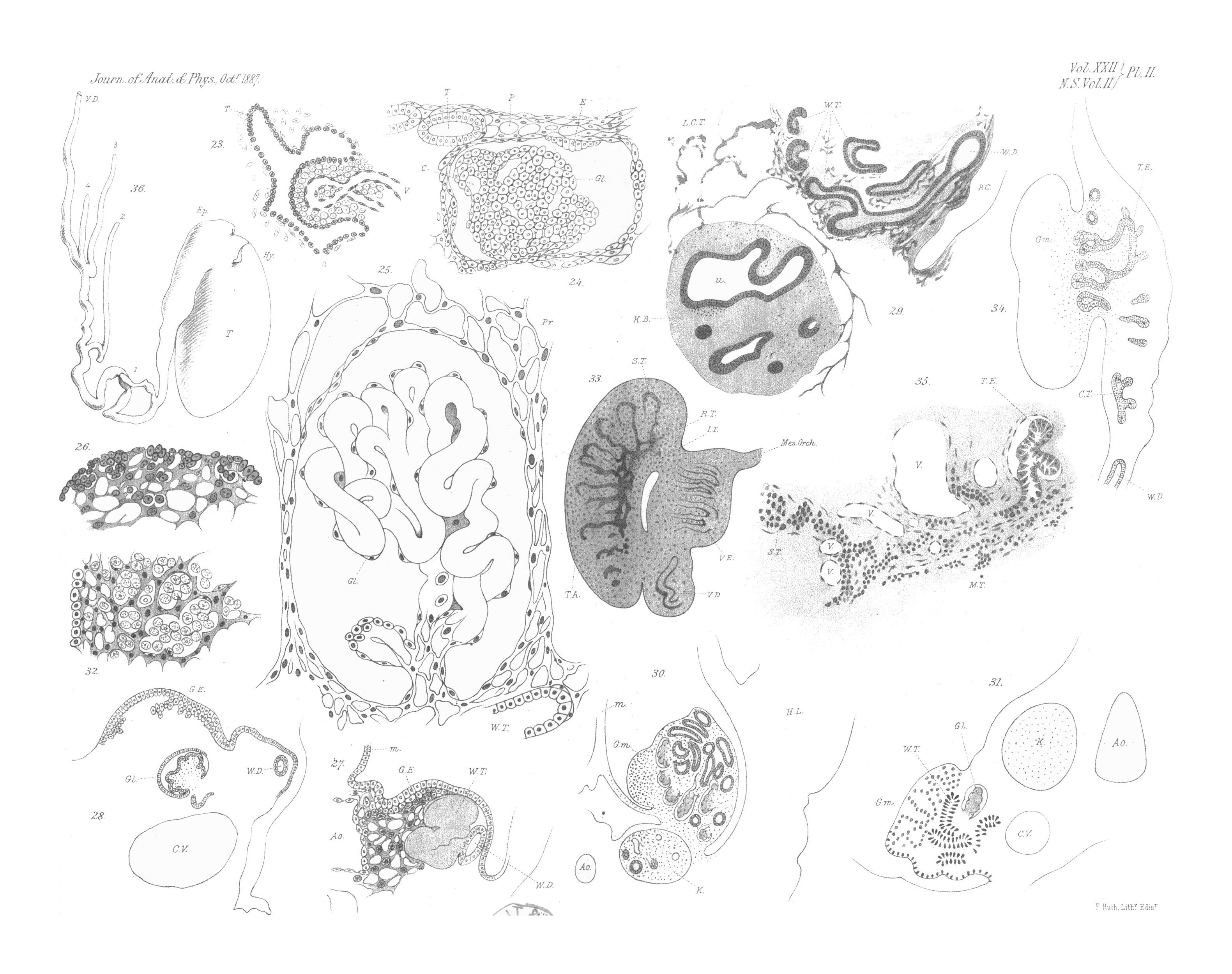 | 3,161 × 2,499 (939 KB) | Z8600021 (talk | contribs) | ==Plate II== ===Reference=== {{Ref-Lockwood1887b}} {{Footer}} |
You cannot overwrite this file.
File usage
The following page uses this file: