File:Lockwood1887b fig33.jpg
Original file (600 × 617 pixels, file size: 101 KB, MIME type: image/jpeg)
Plate II
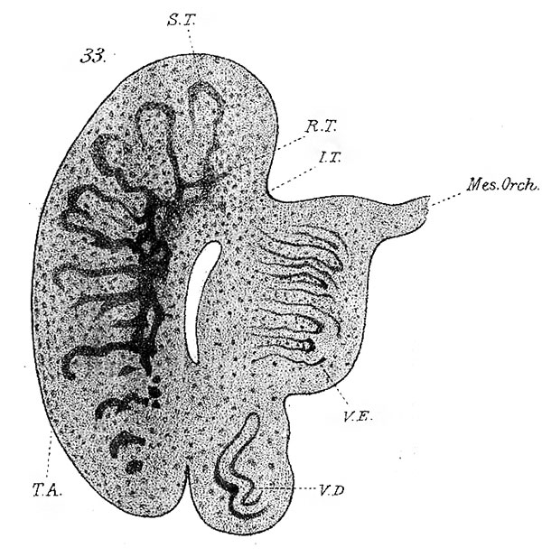
Fig. 33. Testicle and epididymis of human embryo, at about the twelfth week of intrauterine life. The section is not quite longitudinal. x 25. SZ, seminal tubules; RT, rete testes; Mesor., mesorchium ; Vas. Def., vasa deferens; VE, vasa efferentia; I7, indifferent tissue ; 7A, tunica albuginea.
Reference
Lockwood CB. Development and transition of the testis, normal and abnormal. (1887) J Anat. 22(1): 38-77. PMID 17231729
Cite this page: Hill, M.A. (2024, April 26) Embryology Lockwood1887b fig33.jpg. Retrieved from https://embryology.med.unsw.edu.au/embryology/index.php/File:Lockwood1887b_fig33.jpg
- © Dr Mark Hill 2024, UNSW Embryology ISBN: 978 0 7334 2609 4 - UNSW CRICOS Provider Code No. 00098G
File history
Click on a date/time to view the file as it appeared at that time.
| Date/Time | Thumbnail | Dimensions | User | Comment | |
|---|---|---|---|---|---|
| current | 18:58, 14 April 2020 | | 600 × 617 (101 KB) | Z8600021 (talk | contribs) |
You cannot overwrite this file.
File usage
The following page uses this file: